
Table of Contents
- The Main Parts of a Mushroom
- The Cap (Pileus)
- Gills, Pores and Teeth: The Hymenophore
- The Stem (Stipe)
- Veils, Rings and the Volva
- The Part You Do Not See: Mycelium
- Where It All Leads: Spores
- Studying Gills and Spores at Home
- How to Record Mushroom Anatomy for Identification
- A Quick Note on Legality
- Sources
- FAQs
The main parts of a mushroom are the cap (pileus), the gills or pores underneath it (the hymenophore), the stem (stipe), and, in some species, veil remnants such as a ring on the stem or a cup-like volva at the base. Below ground sits the mycelium, the network of threads that actually makes up most of the fungus. The mushroom itself is just the fruiting body, built to release spores.
- A mushroom is the fruiting body of a fungus, and all the parts of a mushroom serve one job: making and releasing spores.
- Gills, pores and teeth all do the same thing: they carry the spore-producing layer, called the hymenium.
- How the gills meet the stem (free, adnate, decurrent and so on) is one of the most useful ID features you can record.
- A ring comes from the partial veil. A volva comes from the universal veil. Not every species has either.
Mushroom Anatomy
The Main Parts of a Mushroom
Ask most people to draw a mushroom and you get a dome on a stick. That sketch is not wrong, but it skips most of what a mycologist actually looks at. Field guides and identification keys lean on a fairly small vocabulary for the parts of a mushroom, and once you know it, species descriptions stop reading like a foreign language.
It also helps to remember what you are looking at. The mushroom is only the reproductive structure, a bit like an apple on a tree. The fungus itself lives in soil, wood or leaf litter as a web of fine threads, and it can stay there for years between flushes of mushrooms.
Here are the parts you will see named again and again:
- Cap (pileus): the top of the mushroom, which shelters the spore-bearing surface.
- Gills (lamellae), pores or teeth: the structures under the cap where spores form.
- Stem (stipe): lifts the cap off the ground so spores can catch air currents.
- Ring (annulus): a skirt or band on the stem, left by the partial veil.
- Volva: a sac or cup at the base, left by the universal veil.
- Mycelium: the thread network in the substrate that the whole thing grows from.
The Cap
The Cap (Pileus)
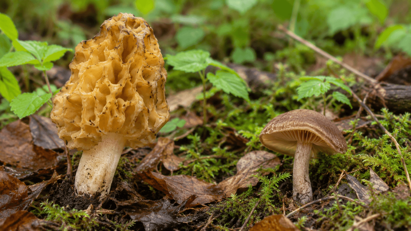
Of all the parts of a mushroom, the cap is usually the first thing you notice and the first thing a key asks about. Mycologists record its shape, size, color, surface texture and whether it changes as the mushroom ages. Many caps start out rounded or bell-shaped and flatten as they open, so it pays to note the age of the specimen too.
The outer skin of the cap is the cuticle, or pileipellis. It can be smooth, sticky, scaly, fibrous or powdery. Under the skin is the flesh, called the context, which may change color when cut or bruised. That is worth writing down, because a color change can separate two otherwise similar species.
Convex
Rounded like an upturned bowl. The most common shape in young caps.
Campanulate
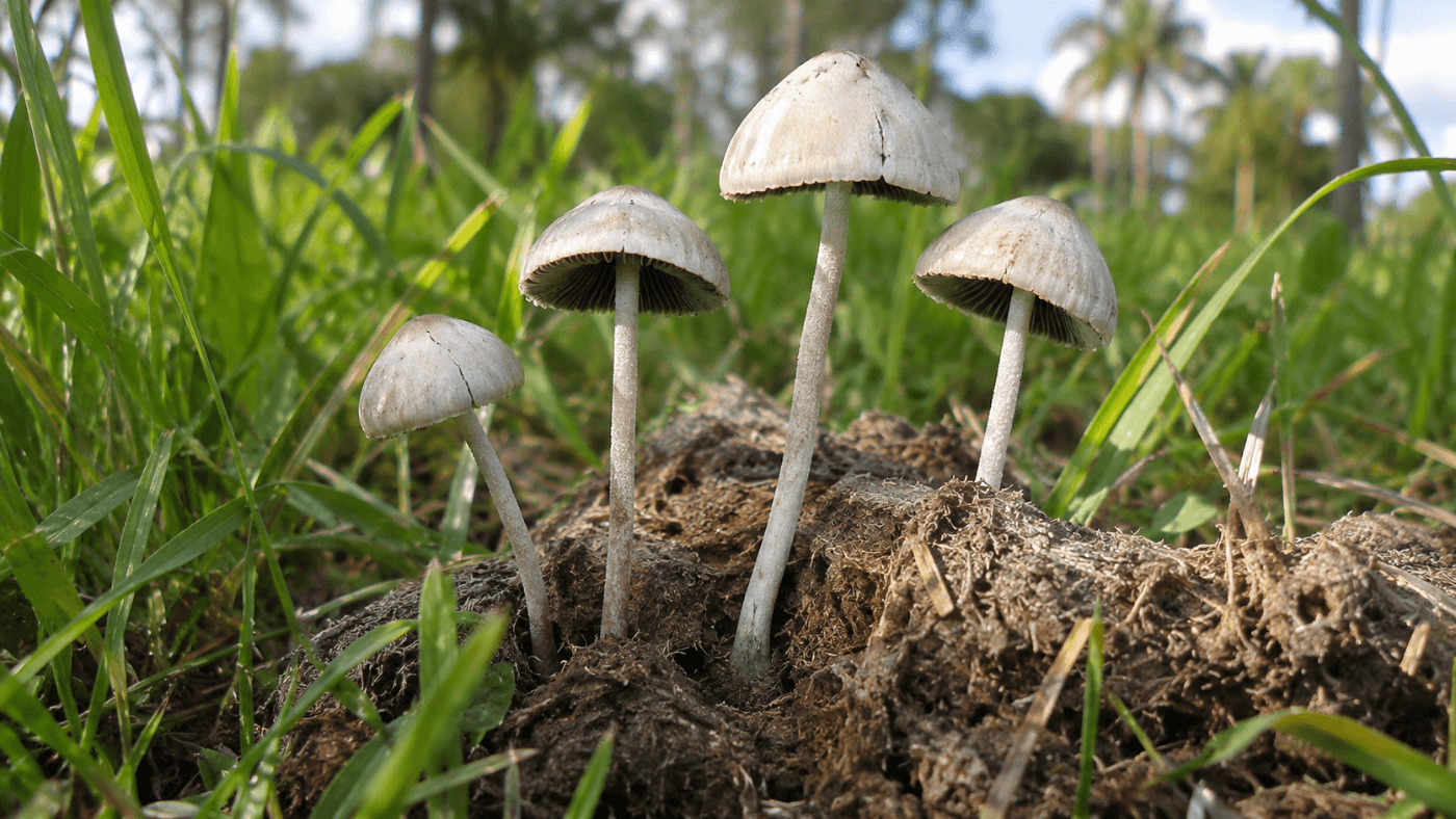
Bell-shaped, taller than wide. Typical of many Panaeolus species.
Umbonate
Flat or convex with a raised bump, the umbo, at the center.
Infundibuliform
Funnel-shaped with a sunken middle, common in chanterelle-like fungi.
The cap margin, the outer edge, has its own terms. It can be striate (lined), incurved, wavy or hung with bits of veil tissue called appendiculate remnants. Small details like these are easy to miss in the field and very easy to miss in a photo, so look at the edge before you move on.
Spore-Bearing Surface
Gills, Pores and Teeth: The Hymenophore
Of all the parts of a mushroom, the underside is the one that matters most to the fungus. The underside of the cap carries the hymenophore, the structure that holds the spore-making layer. In gilled mushrooms these are thin blade-like plates called lamellae. In boletes and polypores they are tiny tubes that open as pores. Hedgehog fungi carry soft spines, or teeth.
All three designs solve the same problem. Folding the hymenium into plates, tubes or spines packs a large surface area into a small cap, which means more spores from one fruiting body. A single large gilled mushroom can release spores in the billions.
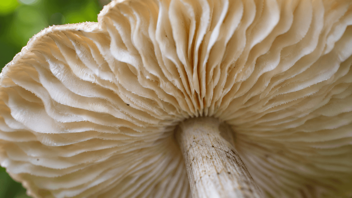
When describing gills, note their color, how crowded they are, whether short gills (lamellulae) sit between the full-length ones, and what the edges look like. The detail people most often skip is attachment, meaning how the gills meet the stem:
| Attachment | What it looks like | Example genera |
|---|---|---|
| Free | Gills stop short and never touch the stem | Agaricus, Amanita, Pluteus |
| Adnexed | Gills touch the stem by a narrow point | Many Panaeolus and Mycena |
| Adnate | Gills meet the stem squarely along most of their width | Psilocybe, Stropharia |
| Sinuate | Gills notch upward just before joining the stem | Tricholoma, Hygrocybe |
| Decurrent | Gills run down the stem below the cap | Pleurotus, Clitocybe |
The easiest way to check attachment is to cut the mushroom in half from top to bottom and look at the profile. Seen from below, adnexed and free gills can look almost the same.
The Stem
The Stem (Stipe)
Among the parts of a mushroom, the stem has the simplest job. It holds the cap up where moving air can carry spores away. Not every mushroom has one. Oyster mushrooms often have a short, off-center stem, and many bracket fungi have none at all and grow straight out of wood.
For identification, note the stem's length and thickness, whether it is centered under the cap, whether it is hollow or solid, and what the base looks like. Some stems are smooth, some are covered in fine hairs or scales, and some stain blue or another color where they are handled. A bulbous base or a base buried in a cup is a big clue, which brings us to veils.
Field tip: Dig out the whole base instead of snapping the stem. A volva often stays underground, and leaving it behind removes one of the most important features in the genus Amanita.
Veils
Veils, Rings and the Volva
Many mushrooms start life wrapped in protective tissue called veils, and they are some of the most overlooked parts of a mushroom. As the mushroom grows, the veils tear, and the pieces left behind are some of the most useful ID features there are. There are two kinds, and they leave very different marks.
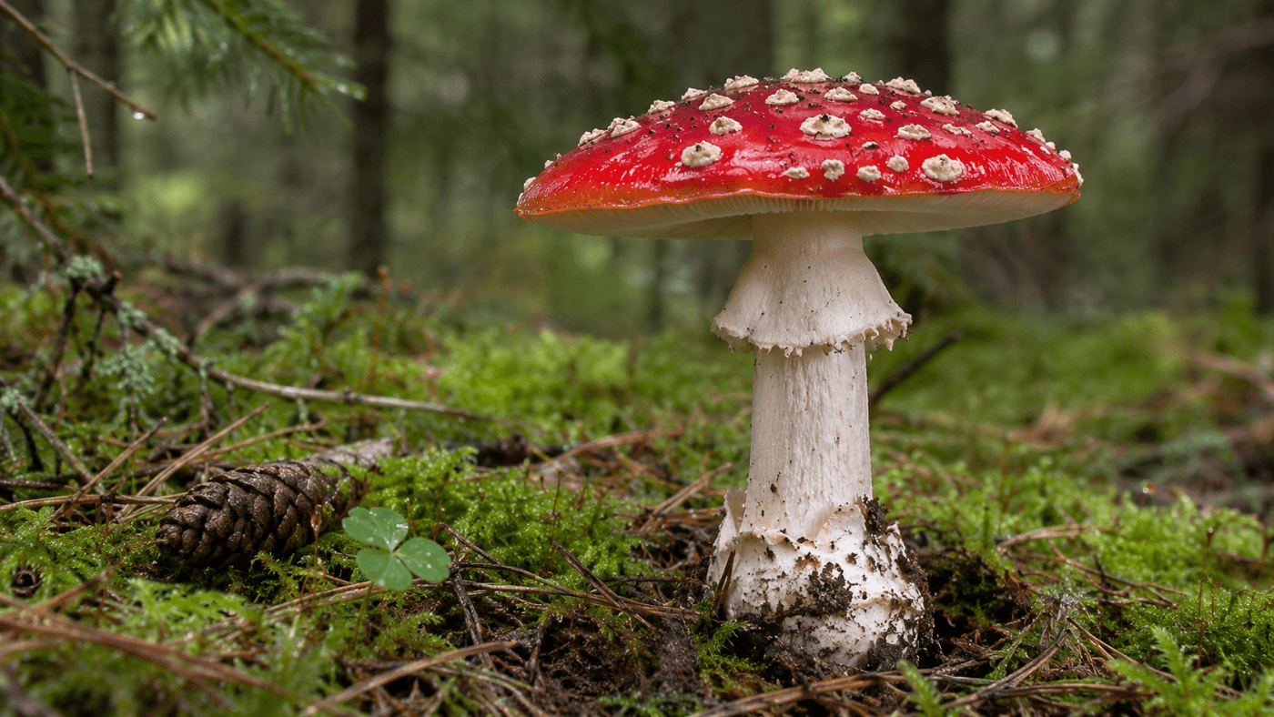
The partial veil stretches from the cap edge to the stem and covers the young gills. When the cap expands, it breaks. What stays on the stem becomes the ring, or annulus. In some genera, such as Cortinarius, the partial veil is a cobweb-like curtain called a cortina, and it leaves only faint fibers on the stem, often dusted with spores.
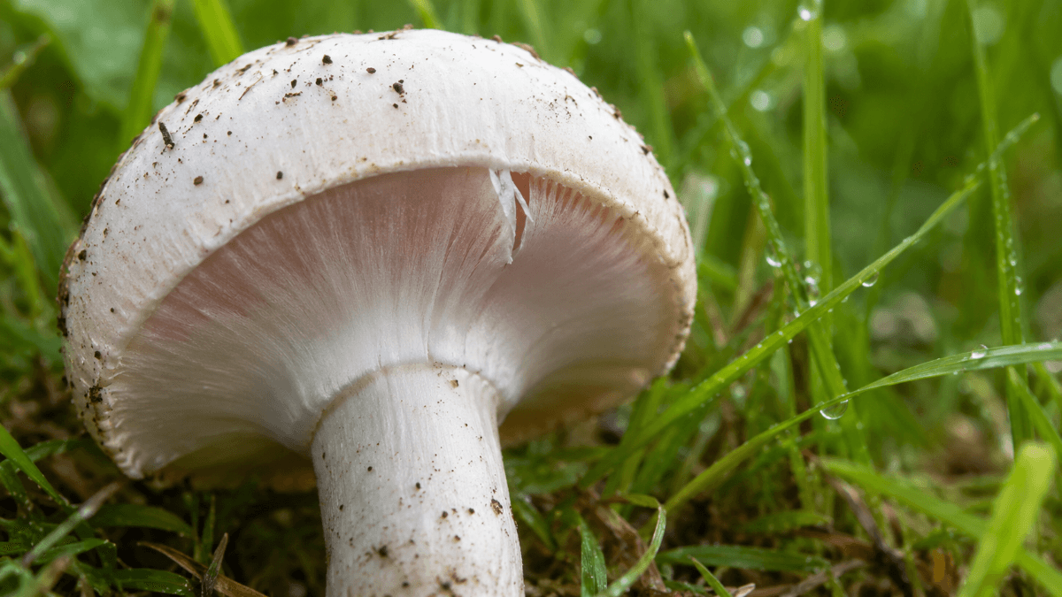
The universal veil wraps the entire young mushroom like an egg. As the mushroom pushes up, this outer layer splits. The bottom part can remain as a sac-like volva at the base, and fragments on top can stay as warts or patches on the cap. The white spots on the classic red fly agaric are universal veil remnants.
Annulus
A ring or skirt on the stem. Comes from the partial veil.
Cortina
A cobweb-like partial veil. Leaves faint fibers, not a true ring.
Volva
A cup or sac at the stem base. Comes from the universal veil.
Warts
Patches on the cap. Also universal veil tissue, and easy to wash off in rain.
Rings can fall off and warts can wash away, so the absence of a veil feature does not prove a species never had one. Check several specimens of different ages when you can.
Below Ground
The Part You Do Not See: Mycelium
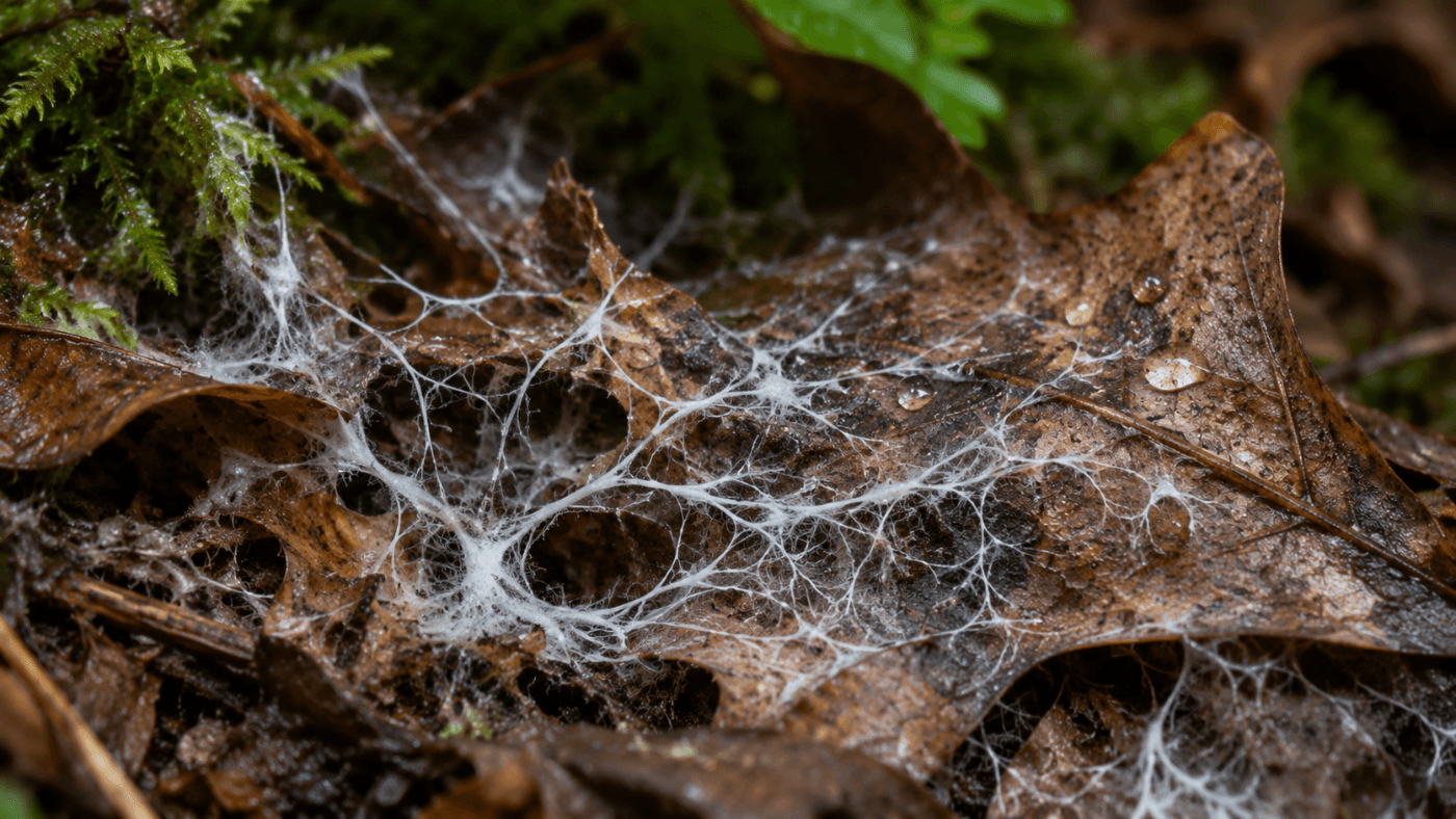
Under every mushroom is the mycelium, a network of microscopic threads called hyphae. It is rarely listed among the parts of a mushroom, but it is the actual body of the fungus. It digests wood, leaves or soil, and only when conditions line up does it put out a fruiting body.
The mushroom you pick up is made of the same threads, packed tightly together. Slice a bit of stem tissue onto a slide and you can see them. Our guide to what hyphae are and how they look under the microscope covers that side of things in detail.
Spores
Where It All Leads: Spores
Every one of the parts of a mushroom covered above exists to get spores made and released. Spores form on microscopic club-shaped cells called basidia in gilled mushrooms, usually four to a basidium. Their color in mass, called the spore print color, is one of the first things a key will ask for, and their size and shape under the microscope can split close relatives.
Spore size varies more than most people expect. The ranges below show typical published spore lengths for a few well-known mushrooms, which is why a calibrated eyepiece reticle is worth having.
Typical Spore Length by Species (µm)
Published length ranges for six familiar mushrooms. Scale: 4–18 µm.
If you want a deeper look at what spores are and how they differ between groups, start with What Is a Spore? and our guide to species ID through spore characteristics.
Reference Material
Studying Gills and Spores at Home
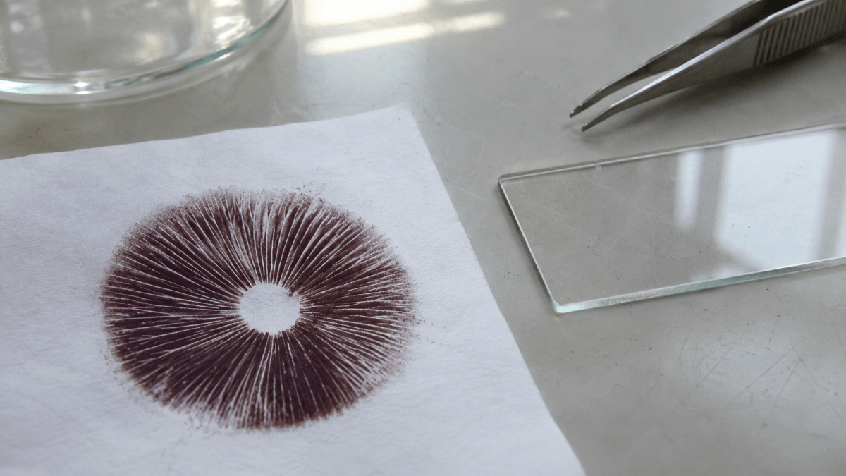
Knowing the parts of a mushroom in the field only gets you so far. A lot of species are separated by what the hymenium produces, and the simplest way to study that is a spore print. Prints keep for a long time when stored dry, and they let you compare spore color and microscopic shape between specimens months apart.
Golden Teacher is a classic starting point for microscopy because its dark purple-brown prints are easy to read and its spores are large enough to measure without much trouble.
Golden Teacher Mushroom Spore Prints
$24.99
$37.48
GT Spore Print The GT Spore Print offers a high-quality sample of the well-regarded Golden Teacher strain, celebrated for its distinct characteristics and long-standing presence in the mycology community. This strain has earned recognition among researchers and taxonomy enthusiasts for… read more

Lab Workflow
How to Record Mushroom Anatomy for Identification
A good description beats a good photo every time. Photos flatten depth and hide the underside, and they almost never show attachment or the base. This is a simple routine for recording the parts of a mushroom for taxonomy work.
- Photograph from three angles. Top of the cap, the underside, and the whole mushroom from the side, with something for scale.
- Measure the cap and stem. Note width, height and thickness in millimeters, plus the age of the specimen.
- Describe the cap surface and margin. Texture, stickiness, color, and any veil remnants at the edge.
- Cut it lengthwise. Check gill attachment in profile, stem hollowness and any color change in the flesh.
- Check the base. Look for a bulb, a volva or a ring, and note where any ring sits on the stem.
- Take a spore print. Lay the cap gills-down on white and black paper or foil for several hours and record the color.
Our microscopy notebook template has fields for all the parts of a mushroom covered above, and the microscope guide covers what you need to measure spores once you have a print.
New to the field? What Is Mycology? is a good primer on where anatomy fits into taxonomy as a whole.
Legal Note
A Quick Note on Legality
Studying the parts of a mushroom and collecting spore prints for microscopy is legal in most of the United States, since most spores contain no scheduled compounds. A few states restrict spores themselves. We do not ship to California, Georgia, Idaho or Florida, and you should check your own local rules before ordering.
Start a Spore Print Reference Set
Golden Teacher prints are a reliable first sample for spore color, shape and size. Every print ships sealed with lot tracking, for microscopy and taxonomy study only.
Golden Teacher Prints All Spore PrintsKeep reading: more guides from our microscopy library.
Sources
- Lamella (mycology), Wikipedia (gill structure and attachment terms).
- Universal veil and Partial veil, Wikipedia (veil types, annulus, volva, cortina).
- Pileus (mycology), Wikipedia (cap shapes and surface terms).
- Stipe (mycology), Wikipedia (stem features and base structures).
- Basidium, Wikipedia (spore formation on basidia).
Magic Spore Labs sells spores and spore prints for microscopy, identification and taxonomy research only. Nothing on this page is guidance on cultivation or consumption. We do not ship to California, Georgia, Idaho or Florida.
FAQs
What are the main parts of a mushroom?
The main parts of a mushroom are the cap (pileus), the gills or pores under the cap, the stem (stipe), and in some species a ring (annulus) on the stem and a volva at the base. The mycelium, a network of fine threads in the soil or wood, is the main body of the fungus.
What are the gills on a mushroom for?
Gills carry the hymenium, the layer of cells that makes spores. Folding that layer into thin plates packs a lot of spore-making surface under a small cap. Pores and teeth do the same job in boletes, polypores and tooth fungi.
What is the ring on a mushroom stem called?
The ring is called the annulus. It is what is left of the partial veil, a thin layer of tissue that covered the young gills and tore away from the cap edge as the mushroom opened.
What is the difference between a partial veil and a universal veil?
A partial veil covers only the gills and runs from the cap edge to the stem, leaving a ring when it breaks. A universal veil wraps the whole young mushroom like an egg and can leave a volva at the base and warts on the cap. Amanita species often show both.
What does gill attachment mean?
Gill attachment describes how the gills meet the stem. Common types are free (not touching the stem), adnexed (touching narrowly), adnate (joined broadly), sinuate (notched) and decurrent (running down the stem). Cutting the mushroom in half lengthwise is the easiest way to check.
Is the mushroom the whole fungus?
No. The mushroom is only the fruiting body, the part that makes and releases spores. Most of the fungus lives out of sight as mycelium, a web of microscopic hyphae growing through soil, wood or leaf litter.