
Table of Contents
Basidiospores are spores that form on the outside of a club-shaped cell called a basidium, usually four at a time, and they belong to the Basidiomycota, the group that includes most gilled mushrooms, boletes and puffballs. Ascospores form inside a sac-like cell called an ascus, usually eight at a time, and they belong to the Ascomycota, which includes morels, cup fungi, truffles and yeasts. Under a microscope, the quickest way to tell them apart is to find where the spores are being made.
- Basidiospores sit on the outside of a basidium on tiny prongs called sterigmata. Ascospores develop inside an ascus.
- Four basidiospores per basidium and eight ascospores per ascus are the usual numbers, though both vary.
- Most basidiospores are shot off with a water droplet trick called Buller's drop. Many ascospores are squirted out of the ascus tip.
- A spore print from a gilled mushroom is a pile of basidiospores. Cup fungi and morels give ascospores.
Spore Basics
What Are Basidiospores?
If you have ever made a spore print from a mushroom cap, you have already collected these spores. They are the sexual spores of the Basidiomycota, one of the two big groups of fungi that make large fruiting bodies. That group covers nearly every mushroom with gills, plus boletes, chanterelles, puffballs, bracket fungi and jelly fungi.
The name comes from the basidium, the cell that makes them. A basidium is a small club-shaped cell lining the gills or pores. Inside it, two nuclei fuse and then divide by meiosis, and the resulting nuclei move up into spores that bud from the top of the cell. Each spore sits on its own thin stalk, called a sterigma.
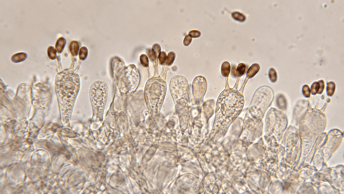
Most basidia carry four spores. Some carry two, as in the cultivated button mushroom, Agaricus bisporus, which is where the name "bisporus" comes from. A few species carry one, three, six or eight.
Where they form
On the outside of a basidium, at the tips of sterigmata.
How many
Usually four per basidium. Two-spored basidia are common in some species.
Who makes them
Basidiomycota: gilled mushrooms, boletes, puffballs, polypores, rusts and smuts.
Telltale detail
A small hilar appendix where the spore was attached to its sterigma.
The Other Group
What Are Ascospores?
Ascospores are the sexual spores of the Ascomycota, the largest phylum of fungi. This group includes morels, cup fungi, truffles, many lichens, most common molds and the yeasts used in baking and brewing. Many ascomycetes never make anything you would call a mushroom, but some do, and those are the ones a mushroom hunter runs into.
Instead of budding off the outside of a cell, ascospores develop inside one. The cell is called an ascus, a sac-like tube packed side by side with other asci in the fertile layer. After meiosis, and usually one extra round of mitosis, the ascus holds eight spores lined up inside it.
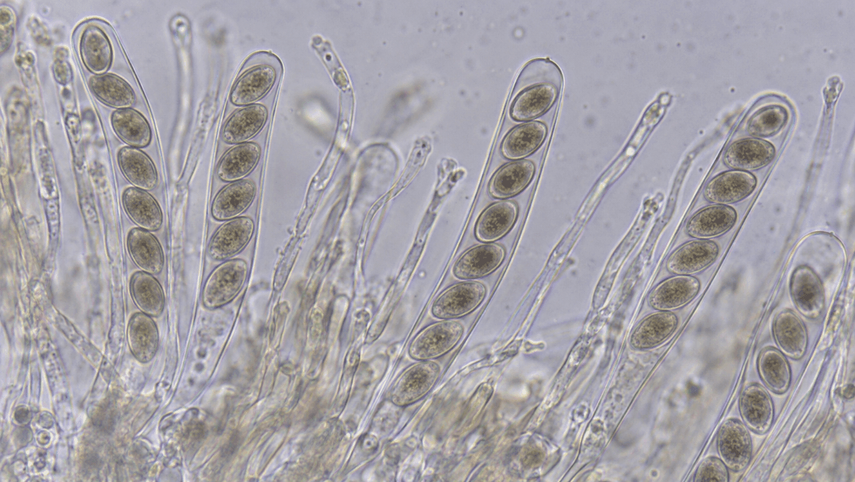
When the ascospores are ripe, pressure builds inside the ascus until it bursts at the tip and fires the spores out. In many cup fungi you can actually see this as a puff of smoke when you breathe on a mature cup. In truffles the asci stay closed, and animals do the dispersal work by digging them up.
Side by Side
Basidiospores vs Ascospores: Key Differences
The two spore types do the same basic job, but nearly everything about how they are made and released is different. Here is how they compare on the points that matter at the microscope:
| Feature | Basidiospores | Ascospores |
|---|---|---|
| Group | Basidiomycota | Ascomycota |
| Where they form | Outside a basidium, on sterigmata | Inside an ascus |
| Usual number | 4 per basidium (2 in some) | 8 per ascus (varies) |
| Release | Shot off by Buller's drop, then fall from gills or pores | Squirted out through the ascus tip, or left for animals |
| Familiar examples | Psilocybe, Agaricus, Amanita, boletes | Morels, Peziza cups, truffles, yeasts |
| What you see in a print | A clean print shaped like the gills | Often a faint dusting, if any |
If you only remember one thing, make it location. Spores on the outside of a club mean basidiospores. Spores sealed inside a tube mean ascospores.
Spore Release
How Basidiospores Launch: Buller's Drop
These spores have one of the neatest launch systems in biology. A tiny droplet of water, called Buller's drop after the mycologist A. H. R. Buller, forms at the base of each spore. At the same time a thin film of water spreads over the spore. When the drop and the film touch and merge, the shift in surface tension flings the spore off its sterigma.
The spore only travels a fraction of a millimeter, just far enough to clear the gill face and drop into the gap between gills. From there it falls out under the cap and rides air currents away. This kind of forcibly shot spore is called a ballistospore.
Why prints work: Because the spores drop straight down from the gills, a cap left on paper overnight lays down a clean copy of the gill pattern. That is why spore prints are so useful for gilled mushrooms and much less useful for most ascomycetes.
Not every basidiomycete uses this system. Puffballs and earthstars hold their spores inside a closed ball and release them as a cloud when raindrops or animals squeeze the case.
At the Microscope
Telling Them Apart Under the Microscope
Loose spores on a slide can be tricky, because shape alone does not always give the group away. A smooth brown ellipse could be a basidiospore from a mushroom or an ascospore from a fungus growing on wood. These are the clues that help:
- A hilar appendix: mushroom spores often show a small nub at one end where they were attached to the sterigma. Ascospores lack it.
- Germ pore: many dark-spored mushrooms, including Psilocybe and Panaeolus, have a flattened pore at the tip of the spore.
- Spores still attached: if you can find spores on basidia or inside asci in a tissue mount, the question answers itself.
- Ornament and septa: some ascospores have cross-walls or warty, netted surfaces. Many mushroom spores are smooth, though Russula and Lactarius spores are famously warty.
Size overlaps a lot between the two groups, but it still helps you narrow things within a genus. The chart below shows typical published spore lengths for a few common species from each group.
Spore Length: Basidiospores vs Ascospores (µm)
Typical published length ranges. Green bars are basidiospores, the black bar is a morel ascospore. Scale: 4–26 µm.
For more on measuring spores and reading their shape, see our guide to species ID through spore characteristics.
Reference Material
Building a Basidiospore Reference Set
The easiest way to get comfortable with this spore type is to look at a lot of them. Prints from different Psilocybe cubensis lines are a good place to start because the spores are large, dark and easy to measure, and you can compare shape and size across isolates on the same slide session.
Keeping a few prints on hand also gives you a known reference when you are working out what a mystery spore is. If the unknown matches the size and germ pore of a known reference, you are probably looking at the same kind of thing.
Mushroom Spore Prints Pick 5 Bundle
$89.96
$99.95
Pick 5 Mushroom Spore Prints Build your Psilocybe cubensis reference collection with the Magic Spore Labs Spore Print Pick 5 Bundle, a curated set of five researcher-selected spore prints, each produced under controlled sterile conditions for reliable microscopy study. Spore… read more

Lab Workflow
How to Observe Basidiospores and Ascospores
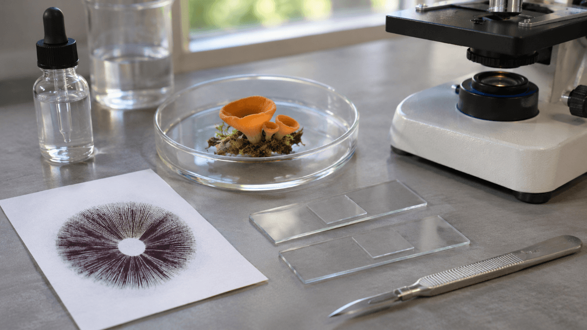
You do not need two separate setups for this. A compound microscope with a 40x objective, an oil immersion lens if you have one, and a few simple mounts will show you both spore types. This is a microscopy-only routine.
- Start with a print. Scrape a tiny amount of spore print onto a clean slide with a sterile blade.
- Add a mounting fluid. Water works for a first look. 3% KOH helps dark spores stand out and softens tissue.
- Find basidia directly. For a gilled mushroom, cut a thin slice across a gill and mount it. Look along the gill edge for club-shaped cells with spores still attached.
- Find asci directly. For a cup fungus, pick a speck of the inner fertile layer, squash it gently and look for long tubes packed with spores.
- Measure 20 spores. Use a calibrated reticle at 400x or 1000x and record length, width and the length-to-width ratio.
- Note the details. Hilar appendix, germ pore, color in KOH and any ornament or septa.
Our microscopy notebook template has columns for every measurement above, and the microscope guide covers which objectives matter for spore work.
If you are newer to this, What Is a Spore? and our guide to the parts of a mushroom give the background for where basidia sit on the gills.
Legal Note
A Quick Note on Legality
Studying mushroom spores under a microscope is legal in most of the United States, and spore prints are sold for exactly that purpose. A few states restrict certain spores. We do not ship to California, Georgia, Idaho or Florida, so check your local rules before ordering.
See Basidiospores for Yourself
Pick any five spore prints and compare their basidiospores side by side at 400x. Every print ships sealed with lot tracking, for microscopy and taxonomy study only.
Spore Prints Pick 5 All Spore PrintsKeep reading: more guides from our microscopy library.
Sources
- Basidiospore, Wikipedia (formation on basidia, sterigmata, hilar appendix).
- Ascospore and Ascus, Wikipedia (eight-spored asci, discharge).
- Ballistospore, Wikipedia (Buller's drop and forcible discharge).
- Basidiomycota and Ascomycota, Wikipedia (group membership and examples).
Magic Spore Labs sells spores and spore prints for microscopy, identification and taxonomy research only. Nothing on this page is guidance on cultivation or consumption. We do not ship to California, Georgia, Idaho or Florida.
FAQs
What are basidiospores?
Basidiospores are the sexual spores of the Basidiomycota, the group that includes most gilled mushrooms, boletes and puffballs. They form on the outside of a club-shaped cell called a basidium, usually four per cell, each on a tiny stalk called a sterigma.
What is the difference between basidiospores and ascospores?
Basidiospores form on the outside of a basidium, usually four at a time, in the Basidiomycota. Ascospores form inside a sac called an ascus, usually eight at a time, in the Ascomycota. Location is the key difference: outside a club versus inside a tube.
How many basidiospores are on a basidium?
Most basidia carry four basidiospores. Some species have two-spored basidia, such as the button mushroom Agaricus bisporus, and a few have one, three, six or eight.
Are morel spores basidiospores?
No. Morels belong to the Ascomycota, so their spores are ascospores. They form in rows of eight inside asci that line the pits of the morel cap, and they are larger than most mushroom spores, often around 18 to 25 micrometers long.
What is Buller's drop?
Buller's drop is a tiny water droplet that forms at the base of a basidiospore. When it merges with a film of water on the spore, the sudden shift in surface tension flings the spore off its sterigma. It is how most mushrooms launch their spores.
Can you see basidiospores with a microscope?
Yes. Most basidiospores are about 5 to 20 micrometers long, so a compound microscope at 400x shows their shape and color, and 1000x oil immersion lets you see details like the germ pore and hilar appendix and measure them accurately.