
Table of Contents
Hyphae are the thin, branching threads that make up the body of a fungus. Each hypha is a tube of cell wall, usually built from chitin, that grows at its tip and is most often 5–10 µm wide. Together, a network of these threads is called a mycelium, and the same threads are packed tightly to form a mushroom's cap, gills and stem.
- A hypha is a single fungal filament. Mycelium is the whole network of them.
- Most fungi have septate hyphae, divided by cross-walls with pores. Many zygomycetes and oomycetes have aseptate (coenocytic) hyphae with no regular cross-walls.
- Clamp connections, small hook-shaped bridges at the septa, are a classic microscope marker of many Basidiomycota.
- Polypores are classified partly by their hyphal system: monomitic, dimitic or trimitic. Turkey tail (Trametes) is the textbook trimitic example.
Fungal Biology
What Are Hyphae?
The word comes from the Greek huphḗ, meaning "web," which is a pretty good description. A hypha is a long, branching filament. In almost every fungus you have heard of, these filaments are how the organism grows, feeds and holds itself together.
Each hypha is basically a tube. The wall is made mostly of chitin, the same tough polymer found in insect shells, which sets fungi apart from plants and from oomycetes (water molds), whose walls are cellulose. Inside the tube is cytoplasm, nuclei and all the usual cell machinery.
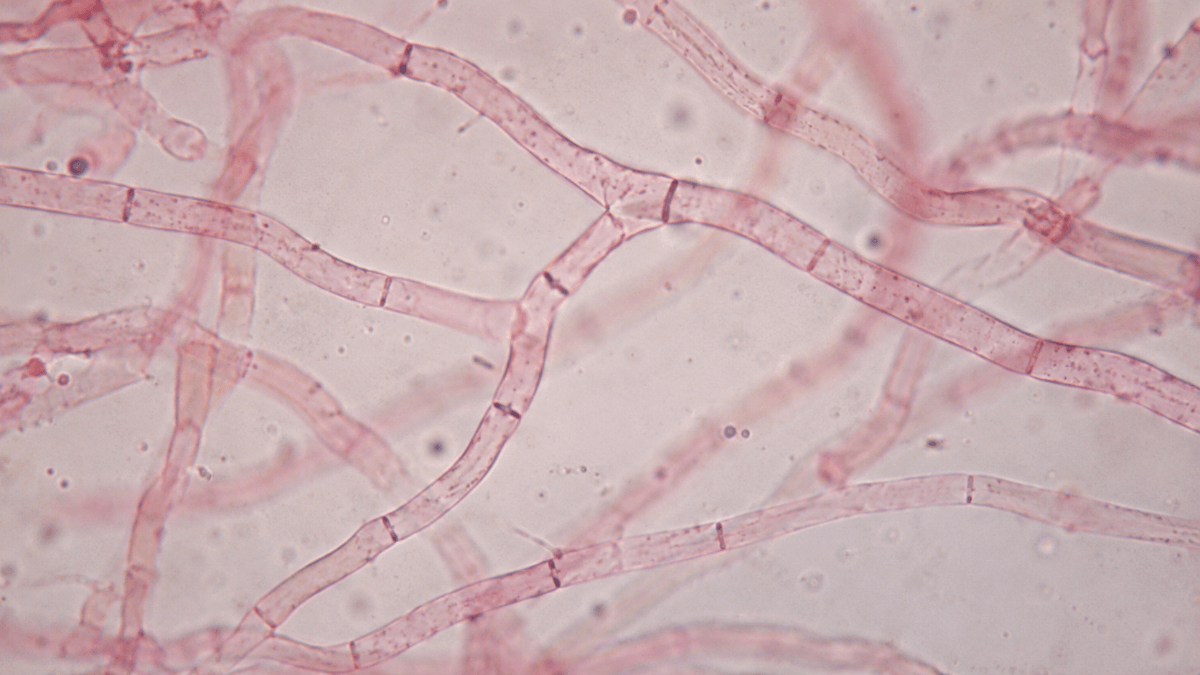
These threads are tiny. Widths run from about 2 µm to 30 µm or more, but most fall in the 5–10 µm range, according to the University of Edinburgh's fungal biology text by Jim Deacon. That is roughly a tenth the width of a human hair, which is why you need a compound microscope to see any real detail.
Wall
Mostly chitin in true fungi. Oomycetes, which look similar, use cellulose instead.
Width
About 2–30 µm, usually 5–10 µm, depending on species and conditions.
Growth
Only at the tip, steered by a cluster of vesicles called the Spitzenkörper.
Network
Branching threads together form the mycelium.
Hyphae vs Mycelium
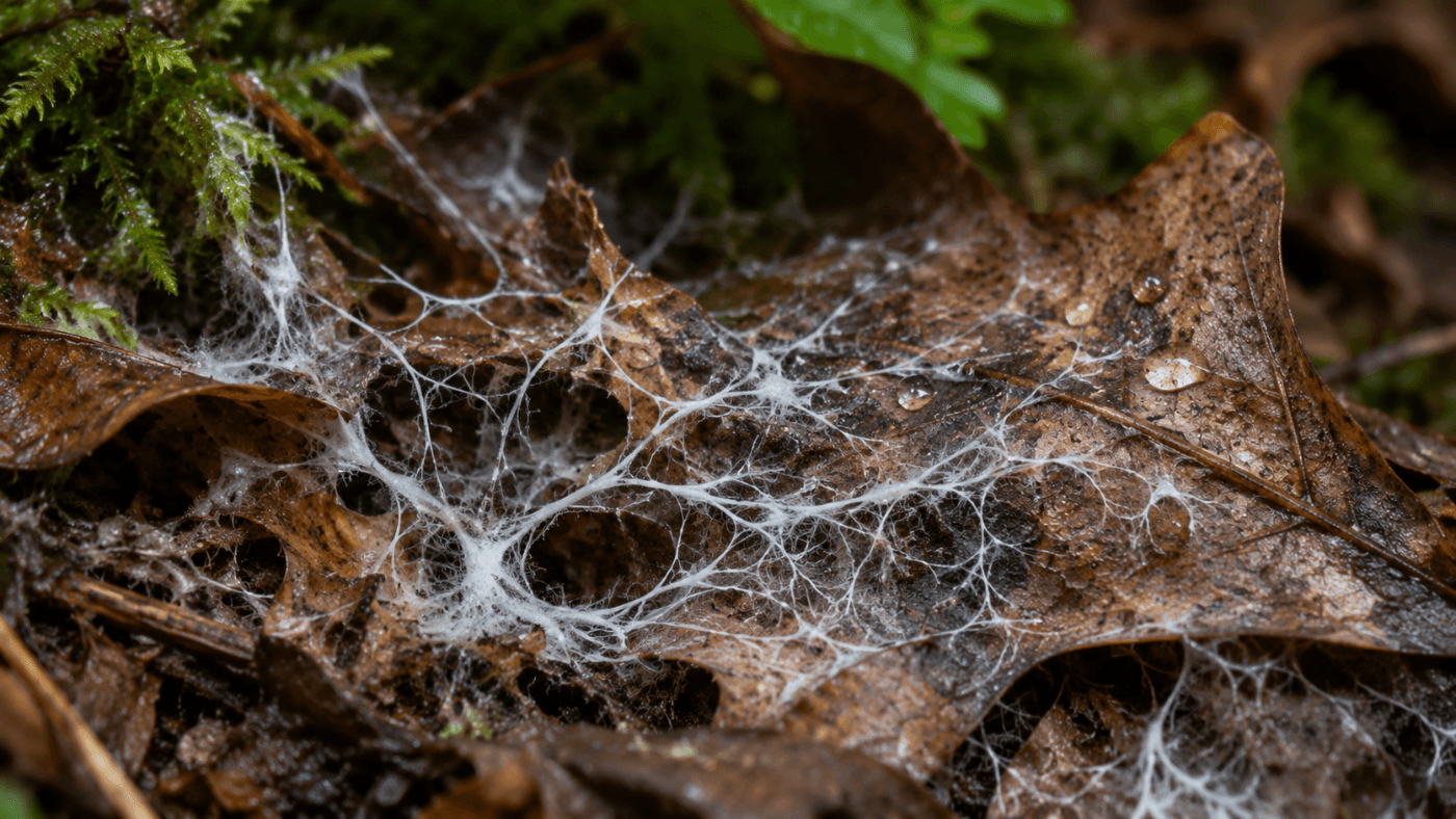
People use the two words interchangeably, but they are not the same thing. A hypha is one thread. Mycelium is the whole mass of interwoven threads, the white, cottony network you see when you flip over a rotting log or a damp pile of leaves.
A mushroom is also made of the same threads, just packed much more tightly. If you tease apart a bit of stem tissue under the microscope, you will find it is a bundle of parallel filaments rather than cells stacked like bricks, which is how plant tissue is built.
Structure
Septate vs Aseptate (Coenocytic) Hyphae
The first thing a mycologist checks on a hypha is whether it has septa. Septa are cross-walls that divide the tube into compartments. They are not solid walls, though. Most have a pore in the middle, so cytoplasm, mitochondria and sometimes even nuclei can move from one compartment to the next.
Fungi without regular septa have aseptate, or coenocytic, filaments: one long continuous tube with many nuclei sharing the same cytoplasm. Here is how the two compare.
| Feature | Septate hyphae | Aseptate (coenocytic) hyphae |
|---|---|---|
| Cross-walls | Regular septa with central pores | None, except to wall off old or reproductive parts |
| Nuclei | One or two per compartment, depending on the fungus | Many nuclei in shared cytoplasm |
| Typical groups | Ascomycota and Basidiomycota (most mushrooms, molds like Aspergillus) | Many Zygomycota (such as Mucor) and Oomycota |
| Seen at 400× | Thin lines crossing the hypha at intervals | A smooth, unbroken tube |
If you are looking at tissue from a gilled mushroom, expect to see septa every time. Mushrooms belong to the Basidiomycota, and their filaments are divided into compartments.
Microscopy
Clamp Connections: A Basidiomycete Fingerprint
Clamp connections are one of the most useful little features in mushroom taxonomy. They show up as a small hook or bump that bridges a septum, curving out from one compartment and fusing with the next.
They form when a hyphal compartment divides. Many basidiomycete filaments carry two different nuclei per compartment, one from each mating partner. The clamp is a side loop that makes sure each new compartment ends up with one of each, keeping that pairing intact as the hypha grows.
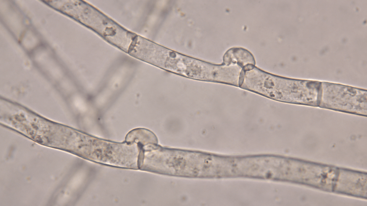
Not every basidiomycete has them, and their presence or absence is often written into species descriptions. That makes clamps a quick yes-or-no character you can record from a simple tissue mount. Ascomycetes do not form clamps, although they have a similar hook structure, called a crozier, in their reproductive tissue.
Where to look: Clamps are easiest to spot at the septa of generative filaments in stem or cap tissue. Scan along a single hypha at 400× and stop at each cross-wall. A clamp looks like a tiny handle on the side of the tube.
Taxonomy
Hyphal Systems in Polypores
Bracket fungi and other polypores can be tough, woody or leathery, and the reason is in their threads. In 1932, E. J. H. Corner described three kinds of filament that show up in these fungi, and the mix of types is still used to sort species today:
- Generative hyphae are thin-walled, often septate, and can carry clamp connections. Every fungus has them, and they are what produce the reproductive structures.
- Skeletal hyphae are long and thick-walled, with few septa and no clamps. They act like rebar and give the fruit body its stiffness.
- Binding hyphae are thick-walled and heavily branched, often looking like deer antlers. They weave the other types together.
A fungus with only the generative type is monomitic, which covers most gilled mushrooms. Add one other type, usually skeletal, and it is dimitic. A fungus with all three is trimitic, and Trametes, the genus that includes turkey tail, is the classic example.
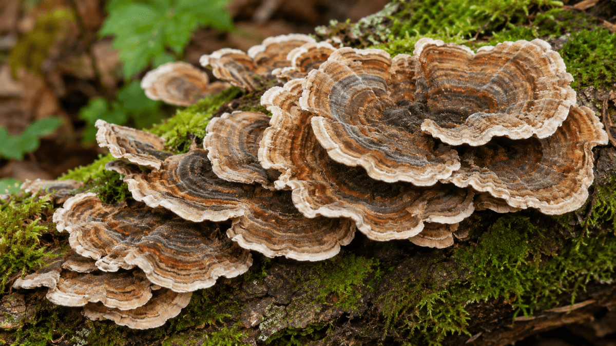
This is why turkey tail feels like thin leather and a gilled mushroom collapses in a day. The difference starts at the level of individual threads, and you can see it for yourself in a crushed tissue mount.
Want to compare hyphal tissue across species? Spore prints are an easy way to start a reference set, since you can pair each print with notes on the fruit body it came from.
Mushroom Spore Prints Pick 5 Bundle
$89.96
$99.95
Pick 5 Mushroom Spore Prints Build your Psilocybe cubensis reference collection with the Magic Spore Labs Spore Print Pick 5 Bundle, a curated set of five researcher-selected spore prints, each produced under controlled sterile conditions for reliable microscopy study. Spore… read more

Specialized Forms
Other Jobs Hyphae Do
Beyond basic growth, these threads take on some surprising shapes depending on how a fungus lives:
- Haustoria, which parasitic fungi push into host cells to absorb nutrients
- Arbuscules, tree-shaped branches inside plant roots formed by mycorrhizal fungi
- Mycelial cords, bundled threads that move water and nutrients over long distances through soil
- Lichen tissue, where fungal threads wrap around algal cells and make up most of the lichen body
- Trapping rings and sticky nets, used by nematode-trapping fungi to catch tiny worms
All of these are still just fungal threads. The basic tube gets modified for a specific job, which is part of why fungi turn up in so many different places.
Lab Workflow
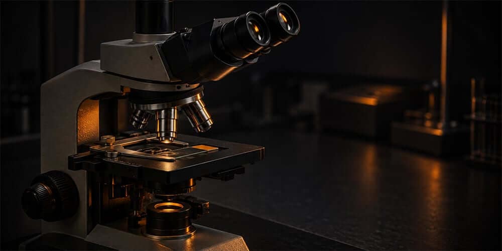
How to Look at Hyphae Under the Microscope
You do not need anything fancy to see them. A compound microscope, slides, a little KOH and some patience will get you septa and, with luck, clamp connections. This is a tissue-observation protocol for microscopy only.
- Take a tiny tissue sample. A sliver the size of a pinhead from a mushroom stem, a gill edge or the inner flesh of a bracket fungus is plenty.
- Mount it in 3% KOH. KOH softens the tissue and helps the threads separate. Add a coverslip.
- Squash gently. Press the coverslip with a pencil eraser to spread the tissue into a thin layer.
- Add a stain if you have one. Congo red or phloxine makes hyphal walls and septa much easier to see.
- Scan at 100×, then 400×. Follow single threads and note septa, branching and wall thickness.
- Check septa at 1000× oil immersion. Look for clamp connections and measure hyphal width with a calibrated micrometer.
Write down the width range, whether clamps are present and any thick-walled threads you see. Our microscopy notebook template has fields for all of it, and our microscope guide covers the objectives you will want.
If you are newer to fungal microscopy, start with the basics in What Is Mycology? and What Is a Spore?. Spores and hyphae are the two structures you will look at most.
Build a Reference Collection for Your Microscope
Pick any five spore prints for side-by-side spore and tissue comparisons. Everything ships sealed with lot tracking, for microscopy and taxonomy study only.
Spore Prints Pick 5 All Spore PrintsKeep reading: more guides from our microscopy library.
Sources
- Hypha, Wikipedia (definition, chitin walls, septa, tip growth and Spitzenkörper, Corner's hyphal systems, specialized hyphae).
- Fungal Biology, Chapter 3: Fungal structure and ultrastructure, J. Deacon, University of Edinburgh (hyphal width, tip extension, aseptate groups).
- Clamp connection, Wikipedia (formation and occurrence in Basidiomycota, croziers in ascomycetes).
- Corner, E. J. H. (1932 and 1966), hyphal system terminology (generative, skeletal and binding hyphae), as summarized in the Wikipedia Hypha article above.
Magic Spore Labs sells spores and spore prints for microscopy, identification and taxonomy research only. Nothing on this page is guidance on cultivation or consumption. We do not ship to California, Georgia, Idaho or Florida.
FAQs
What are hyphae in simple terms?
Hyphae are the microscopic threads that make up a fungus. Each one is a thin tube of cell wall, usually made of chitin, that grows at its tip and branches as it goes. A mass of hyphae is called mycelium, and mushrooms are made of tightly packed hyphae too.
What is the difference between hyphae and mycelium?
A hypha is a single fungal thread. Mycelium is the whole network of interwoven hyphae, like the white cottony mass you see under a rotting log. Think of hyphae as individual strands and mycelium as the whole tangle.
What is the difference between septate and aseptate hyphae?
Septate hyphae are divided into compartments by cross-walls called septa, which usually have a pore in the middle. Aseptate, or coenocytic, hyphae have no regular cross-walls, so many nuclei share one continuous tube. Mushrooms and most molds are septate, while many zygomycetes such as Mucor are aseptate.
What is a clamp connection on a hypha?
A clamp connection is a small hook-shaped bridge that forms at a septum when a dikaryotic hypha divides. It keeps two compatible nuclei paired in each new cell. Clamps are found only in basidiomycetes, so spotting them under the microscope tells you the fungus belongs to that group.
How wide are fungal hyphae?
Most fungal hyphae are about 2 to 30 micrometers wide, and many common species fall between 5 and 10 micrometers. That is far thinner than a human hair, so you need a compound microscope at 400x to see their walls, septa and clamp connections clearly.
Do spores grow into hyphae?
Yes. In biology, a fungal spore germinates by pushing out a germ tube, and that tube becomes the first hypha. For microscopy work, spore prints are studied as-is: you look at spore shape, size and color on a slide rather than germinating them.