
Table of Contents
- What Is a Spore Syringe?
- What Is a Spore Print?
- How Do They Compare for Microscopy Use?
- What Do Spores Look Like Under the Microscope Regardless of Format?
- Which Format Is Better for Long-Term Archival and Taxonomy Reference?
- What About Liquid Culture — How Is It Different from Both?
- Legal Context for Researchers
- FAQs
Quick Answer
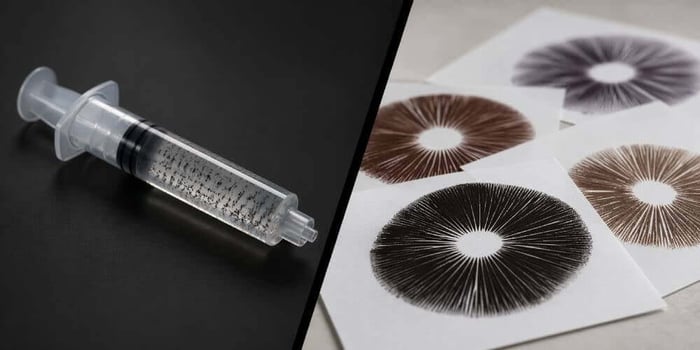
A spore syringe is a sterile aqueous suspension of mushroom spores in a pre-filled syringe — optimized for immediate slide preparation and beginner microscopy. A spore print is a dry deposit of spores on foil or paper — optimized for long-term archival storage and taxonomic reference. Neither is universally superior; the right format depends on your research objectives, experience level, and how you plan to use and store your collection.
Key Takeaways
- A spore syringe suspends spores in sterile distilled water, ready for direct application to a microscope slide with no preparation steps
- A spore print deposits dry spores onto foil or paper from a mature mushroom cap — the preferred long-term archival format
- Spore syringes offer superior ease of use for slide preparation; spore prints offer longer viability and higher spore density per unit
- Under the microscope, both formats reveal identical spore morphology — format does not change what you observe
- Shelf life: syringes 6–12 months refrigerated; prints potentially years to decades when sealed, cool, and dry
- P. cubensis spores contain no psilocybin or psilocin; possession for microscopy research is legal in most U.S. states, with exceptions in California, Georgia, Idaho, and Florida
What Is a Spore Syringe?
A spore syringe is a sterile, pre-filled syringe containing mushroom spores suspended in distilled or sterile water. Mushroom spores are evenly distributed throughout the liquid solution, making them immediately accessible for microscopy without any preparation beyond shaking to redistribute settled spores and applying a drop to a slide.
Standard spore syringes are typically 10mL in volume and contain millions of individual spores from a single strain. The liquid medium keeps spores in a hydrated, mobile state — easy to dispense in controlled volumes, easy to distribute across a slide surface, and easy to handle without specialized equipment or sterile technique beyond what the syringe itself provides.
For the working microscopist, the syringe format is the practical default. There is no rehydration step, no risk of clumping from improperly dried material, and no requirement for additional sterile tools. This is the primary reason spore syringes are recommended as the entry point for anyone beginning to study fungal spore morphology.
Magic Spore Labs spore syringes are prepared in HEPA-filtered environments using sterile distilled water, ensuring each syringe arrives uncontaminated and ready for immediate use. For a step-by-step guide to slide preparation with liquid spores, see our post How to Use Golden Teacher Liquid Spores.
Golden Teacher Mushroom Spore Syringe

$22.99
$24.99
Golden Teacher Liquid Spores Revered as the community's most popular strain for its hallucinogenic properties and mycologists' top choice for its versatility, the Golden Teacher Mushroom Spores strain is undeniably a favorite for many. It's the most common strain of the… read more
What Is a Spore Print?
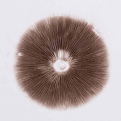
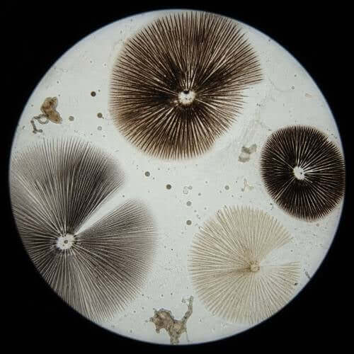
A spore print like the Golden Halo Mushroom Spore Prints is created when a mature mushroom cap is placed gill-side down on a flat sterile surface — typically aluminum foil, mylar, or paper — and left undisturbed while spores fall naturally from the gills onto the surface below. Over several hours, millions to potentially hundreds of millions of spores accumulate in a pattern reflecting the gill layout of the cap.
Once complete, the print is allowed to dry fully and then sealed — typically folded in foil and stored in an airtight container. In this dried, sealed state, a spore print can remain viable for years and, under optimal conditions, for decades.
The color of the print is one of its most scientifically significant features: spore print color is a primary taxonomic diagnostic for identifying mushroom species, and it is only visible in bulk deposit form. Individual spores under the microscope may appear transparent or lightly pigmented — it is the mass of deposited spores that produces the characteristic color visible to the naked eye. For Psilocybe cubensis strains, the characteristic print color is dark purplish-brown to near-black. For other species, colors range from white (Agaricus species) to rust-brown (Cortinarius species) to pink (Pluteus species) — each a meaningful taxonomic data point.
A single spore print contains sufficient material to prepare between 5 and 100 standard 10mL spore syringes, making it an economical and information-dense archival format. The tradeoff is preparation complexity: converting a print to a slide-ready format requires sterile water, clean tools, and care to avoid introducing contaminants during rehydration.
How Do They Compare for Microscopy Use?
Both formats deliver the same result on a prepared slide: mushroom spores ready for examination under magnification. The differences are entirely about preparation workflow, storage, and research context — not about what you observe under the lens.
| Factor | Spore Syringe | Spore Print |
|---|---|---|
| Slide preparation | Direct — apply one drop to slide | Requires rehydration in sterile water first |
| Ease of use | Beginner-friendly | Requires basic sterile technique |
| Spore density per unit | Millions in 10mL suspension | Millions to hundreds of millions in deposit |
| Shelf life | 6–12 months refrigerated | Years to decades sealed, cool, dry |
| Taxonomic color data | Not visible in suspension | Bulk print color is a primary diagnostic |
| Storage requirements | Refrigerate at 2–8°C | Cool, dark, airtight container |
| Cost per observation | Moderate | Lower over time (more uses per unit) |
| Contamination risk | Low when sealed and refrigerated | Low when sealed; higher during rehydration |
| Best suited for | Beginners, regular sessions, convenience | Long-term archival, taxonomic reference collections |
What Do Spores Look Like Under the Microscope Regardless of Format?
This is worth stating clearly: format does not change spore morphology. Whether you prepare a slide from a syringe drop or from rehydrated print material, you are observing the same biological structures.
For Psilocybe cubensis strains — including Golden Teacher, Jedi Mind Fuck, B+, and others in the MSL catalog — the spore features you will observe are consistent across both formats:
- Shape: Subellipsoid — oval form with slightly tapered poles
- Size: Approximately 11–17 × 7–12 μm (strain-dependent; see individual species profiles for precise measurements)
- Germ pore: A clearly visible apical germ pore at one pole, resolvable at 400× magnification
- Wall texture: Smooth, moderately thick-walled — no surface ornamentation
- Color under transmitted light: Individual spores appear pale amber to yellow-brown; bulk print deposits appear dark purplish-brown
At 400× (40× objective + 10× eyepiece), you can reliably observe shape, germ pore, and general dimensions. For detailed wall structure and surface features, 1000× with oil immersion is recommended.
The practical difference between formats at the microscope level is spore density on the slide. A syringe drop disperses spores evenly in suspension, producing a predictable distribution. A rehydrated print suspension can produce variable density depending on how thoroughly the material was mixed — beginners sometimes over-concentrate, resulting in clumped spores that are harder to measure individually. Starting with a syringe eliminates this variable entirely.
Which Format Is Better for Long-Term Archival and Taxonomy Reference?
For building a reference collection intended for comparative taxonomy work over months or years, spore prints are the superior archival format. Three reasons explain this:
Viability longevity
Spores in aqueous suspension degrade gradually even under refrigeration. Dry prints are metabolically inert when sealed — no active biological process is occurring that reduces viability. Well-documented field accounts describe viable prints decades after collection.
Taxonomic color data
Spore print color is a primary species-level diagnostic that is only visible in bulk deposit form. A researcher building a comparative taxonomy collection needs this data preserved — only the print format provides it.
Information density
A single print provides enough material for many microscopy sessions over an extended research period. A syringe, once partially used, begins to degrade the remaining suspension even under refrigeration.
Many experienced microscopists maintain both formats: syringes as their working daily-use tools, and prints as the archival master record. This hybrid approach gives you the best of both — low-friction access for regular observations alongside a long-term reference collection that preserves color data and genetic material indefinitely.
What About Liquid Culture — How Is It Different from Both?
Since this comparison frequently raises the question, it is worth distinguishing liquid culture from both formats above, because it is a fundamentally different biological material.
| Format | Contents | Biological State | Microscopy Use |
|---|---|---|---|
| Spore syringe | Dormant spores in sterile water | Pre-germination — inert reproductive cells | Direct slide preparation |
| Spore print | Dormant spores in dry deposit | Pre-germination — inert reproductive cells | Rehydrate, then slide preparation |
| Liquid culture | Live mycelium in nutritive liquid | Post-germination — active hyphal growth | Not a microscopy research product |
A spore syringe and spore print both contain dormant, ungerminated spores — the reproductive cells of the mushroom, containing no scheduled compounds and appropriate for microscopy research. A liquid culture contains live mycelium: actively growing hyphal tissue in a nutritive liquid medium, representing the next stage of the fungal life cycle after germination. These are biologically, practically, and legally distinct products. Magic Spore Labs sells spore syringes, liquid spores, and spore prints strictly for microscopy and taxonomy research purposes.
Legal Context for Researchers
Psilocybe cubensis spores — in both syringe and print format — do not contain psilocybin or psilocin. At the federal level, the DEA’s position is that ungerminated spores are not controlled substances because they do not contain scheduled compounds. State-level exceptions apply in four states:
| State | Possession Status | Notes |
|---|---|---|
| California | Restricted | Prohibited under state law |
| Georgia | Prohibited | No sales or shipment to Georgia |
| Idaho | Prohibited | No sales or shipment to Idaho |
| Florida | Restricted | Possession restrictions apply |
| All other U.S. states | Legal for microscopy & research | Spores contain no scheduled compounds; cultivation remains federally prohibited regardless of state |
All spore products at Magic Spore Labs are sold strictly for microscopy, taxonomy, and research purposes. Customers are responsible for verifying applicable laws in their jurisdiction prior to purchase.
Build your microscopy collection
Magic Spore Labs carries spore syringes, liquid spores, and spore prints prepared under sterile conditions for microscopy and taxonomy research.
Shop Spore Syringes Shop Spore PrintsDisclaimer: All spores offered by Magic Spore Labs are intended for microscopy, taxonomy, and research purposes only. Spores are not intended for cultivation. It is the customer’s responsibility to know and comply with all local, state, and federal laws regarding possession and use of spore products.
FAQs
What is the difference between a spore syringe and a spore print?
A spore syringe suspends dried spores in sterile distilled water in a pre-filled syringe, ready for immediate slide preparation. A spore print is a dry deposit of spores on foil or paper, created by allowing a mature mushroom cap to release spores naturally onto a surface. Both contain the same spore morphology; the differences are in preparation workflow, shelf life, and research use case.
Which is better for beginner microscopists — spore syringe or spore print?
A spore syringe is the recommended starting format for beginners. It requires no preparation, no sterile technique, and no additional tools — apply one drop to a slide and observe. Spore prints require rehydration in sterile water and careful technique to avoid clumping or contamination during the process.
Do spore format differences affect what you see under the microscope?
No. Spore morphology — shape, size, germ pore, wall texture — is identical regardless of whether spores came from a syringe or a rehydrated print. Format affects how you prepare the slide, not what the spores look like.
How long do spore syringes and spore prints last?
Spore syringes are best used within 6–12 months when refrigerated at 2–8°C. Spore prints sealed in foil and stored cool, dark, and dry can remain viable for years and potentially decades.
What is the difference between a spore syringe and liquid culture?
A spore syringe contains dormant, ungerminated spores in sterile water — pre-germination reproductive cells with no scheduled compounds. A liquid culture contains live mycelium: active, post-germination hyphal growth in a nutritive liquid medium. These are biologically and legally distinct products. Spore syringes and prints are the formats used for microscopy research.
Can a spore print be converted into a spore syringe?
Yes — a single spore print contains enough material to produce between 5 and 100 standard 10mL spore syringes. This conversion requires sterile distilled water, clean tools, and a controlled environment to avoid contamination during the rehydration process.
Is spore print color scientifically meaningful?
Yes. Spore print color is a primary taxonomic diagnostic used to distinguish mushroom species. It is only visible in bulk deposit form — individual spores under the microscope may appear pale or transparent. For Psilocybe cubensis strains, the characteristic print color is dark purplish-brown to near-black. Other species produce prints ranging from white to rust to pink, each carrying taxonomic significance.