

Table of Contents
- Why Golden Teacher Continues to Stand Out
- Origins and Reputation of Golden Teacher
- Observing Golden Teacher Spores Under the Microscope
- Structural Traits That Make Golden Teacher Distinct
- Mycelial Characteristics Under Magnification
- Developmental Traits Documented in Microscopy
- Why Golden Teacher Is a Microscopy Favorite
- Tips for Microscoping Golden Teacher Spores
- Notable Traits Researchers Frequently Document
- Common Questions New Microscopists Ask About Golden Teacher
- Documenting Observations for Research
- Final Thoughts: A Strain That Teaches Through Clarity
- FAQs
Why Golden Teacher Continues to Stand Out
Among all the spore varieties studied in microscopy, few attract as much interest as Golden Teacher. Even when viewed purely through an observational, research-based lens, this strain consistently draws attention for its recognizable appearance, reliable structure, and approachable morphology. Researchers appreciate how its spores present clean, distinguishable traits under magnification, making it a favorite for both beginners and experienced microscopists.
This guide takes a closer look at what makes Golden Teacher so notable when placed under the microscope—its spore characteristics, structural features, developmental patterns, and the unique traits that microscopists often document. The goal is to paint a clear picture of how this strain behaves visually without discussing cultivation or effects. Everything here focuses strictly on microscopy, morphology, and scientific observation.
Origins and Reputation of Golden Teacher
The Story Behind Its Name
Golden Teacher is instantly recognizable by its golden-tinted cap in nature, but under a microscope, its identity is revealed through structural traits rather than coloration. Microscopists often describe the strain as a great “teacher” because of how clearly its spores display hallmark characteristics common to similar species. Its predictable structure makes it helpful for learning the fundamentals of spore identification.
Why Researchers Gravitate Toward It
One of the reasons Golden Teacher remains widely studied is its consistency. Whether examining fresh spore deposits, slides prepared from spore prints, mushroom liquid spores or suspended samples from sterile water solutions, the morphology tends to remain uniform. This reliability makes it an excellent reference point for building observational skill and comparing different spore samples side by side.

Observing Golden Teacher Spores Under the Microscope
General Spore Shape and Size
When placed under magnification, Golden Teacher spores typically appear elliptical, symmetrical, and smooth. Most fall within the size range commonly documented for this species group, and their uniformity helps researchers compare variations across other strains. On a slide, the spores often align in ways that highlight their bilateral shape, allowing for clear recognition and classification.
Coloration and Texture in Microscopy
Unlike the golden hues associated with the fruiting body, the spores tend to display a deep purplish-brown tone when collected in prints. Under brightfield microscopy, they appear darker and more opaque, sometimes revealing subtle gradients depending on their maturity. Researchers also note the smooth exterior texture, which contrasts with the ornamented spores sometimes found in other fungal genera.
Structural Traits That Make Golden Teacher Distinct
Germination Features and Spore Wall Layers
Golden Teacher spores exhibit a firm, clearly defined wall that becomes easier to differentiate at higher magnifications. The dual-layered structure often shows a darker outer outline with a slightly lighter interior. Many microscopists focus on the germination pore—an identifiable feature that appears as a slight indentation or thinning at one end of the spore. Its appearance helps place the strain within broader taxonomic categories during microscopy sessions.
Spore Arrangement Patterns on Prints
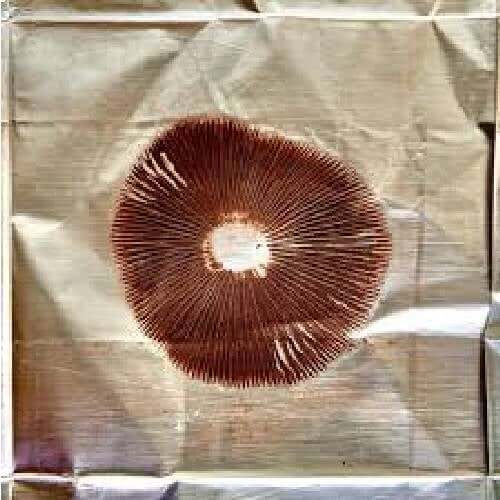
When viewed from properly prepared spore prints, Golden Teacher often produces a dense, even distribution. Under the slide, this can translate into organized clusters rather than chaotic scattering. The spores may group into small patches or align in thin linear formations, depending on how they were transferred onto the glass. These arrangement patterns can be useful when comparing preparation methods or spore collection techniques.
Golden Teacher Mushroom Spore Syringe

$22.99
$24.99
Golden Teacher Liquid Spores Revered as the community's most popular strain for its hallucinogenic properties and mycologists' top choice for its versatility, the Golden Teacher Mushroom Spores strain is undeniably a favorite for many. It's the most common strain of the… read more
Mycelial Characteristics Under Magnification
Hyphal Texture and Branching
If mycelial samples are observed strictly in a microscopy-approved setting, researchers frequently note that Golden Teacher hyphae present a consistent thickness with steady branching angles. The hyphae tend to show smooth walls and predictable septa placement. This uniformity allows microscopists to compare hyphal development across other spore samples with ease and refine their understanding of fungal structure.
Rhizomorphic vs. Cottony Growth Forms
Some microscopy enthusiasts focus on the contrast between rhizomorphic strands—those rope-like structures—and softer cottony segments. Golden Teacher can display both forms, depending on the environmental variables used purely for observational research. Under the lens, rhizomorphic structures may appear more defined, showing thicker hyphal bundles, while cottony growth appears softer and more diffuse, with less obvious directional growth.
Developmental Traits Documented in Microscopy
Early Morphological Indicators
Microscopists who observe spore hydration or early development stages note that Golden Teacher often displays clear initial swelling before other structural changes occur. The germ pore may become more visible, and the inner contents shift slightly as the sample interacts with moisture or mounting mediums. Although these observations remain purely structural, they help teach beginners how to identify early-stage changes without venturing into cultivation discussions.
Mature Structural Patterns
With time, Golden Teacher samples observed through microscopy may develop increasingly defined cellular boundaries. Hyphal sections show thickened walls, and branching may become more frequent and organized. These traits make the strain particularly useful for comparing early versus later stages of morphological development and understanding how fungal structures become more complex over time.
Why Golden Teacher Is a Microscopy Favorite
Consistency Across Slides
One major advantage of observing Golden Teacher is the repeatability of what you see. Slide after slide, its spores display the same elliptical shape, defined germination pore, and smooth exterior. Consistency matters because it allows researchers to confirm their microscopy methods, validate their slide preparation techniques, and trust that unexpected variations likely come from preparation differences rather than the spores themselves.
Great for Beginners and Advanced Users
Beginners appreciate that Golden Teacher spores are easy to recognize. Advanced researchers appreciate how nuanced the small variations can be—color intensity, pore visibility, wall thickness, arrangement patterns, and internal gradients. In this way, the strain provides a wide learning spectrum. It can introduce someone to basic spore morphology while still offering enough detail for deeper, long-term study.
Tips for Microscoping Golden Teacher Spores
Preparing Clear Slides
Quality microscopy begins with clean slides and properly transferred spores. Many researchers lightly scrape the spore print or use a sterile swab to create a thin smear.
Too dense a sample can hide fine details, while too light a sample may not show enough spores to analyze. Golden Teacher’s uniformity helps researchers quickly find the ideal concentration for clear, readable slides.Lighting and Magnification Adjustments
Different magnification levels reveal different traits. Lower magnification highlights arrangement patterns and density, while higher levels reveal pore structure, wall layers, and internal granularity.
Adjusting brightfield intensity or adding extra contrast can make subtle features such as inner vs. outer wall differences or the germination pore outline more visible.
Notable Traits Researchers Frequently Document
Elliptical Symmetry and Defined Pore
Across countless microscopy notes, one trait shows up consistently: symmetry. Golden Teacher spores rarely deviate from their general elliptical form, and the germination pore is often cleanly visible. These consistent traits help reduce confusion when differentiating between similar spore samples and provide a solid reference point for training the eye to spot slight differences in other strains.
Predictable Hyphal Formation
Researchers also document how predictable the hyphal formation appears when observed under controlled research settings. The hyphae tend to maintain a readable pattern of septation, branching, and wall thickness. This makes Golden Teacher ideal for training observational accuracy, as subtle variations in hyphal direction, thickness, or spacing become easier to spot against such a reliable baseline.
Common Questions New Microscopists Ask About Golden Teacher
“Why do Golden Teacher spores look darker under the microscope?”
The darker appearance comes from the natural pigmentation of the spores. Under brightfield light, this pigmentation can cause the spores to absorb more illumination, creating a deeper tone and high contrast against the slide background. This contrast helps highlight their outlines and structural features during observation.
“Are Golden Teacher spores easier to view than other varieties?”
Many researchers say yes—not because the spores are dramatically larger or more unusual, but because their symmetry, pore clarity, and coloration make them visually straightforward to recognize. This clarity simplifies the learning process for people who are still getting comfortable with focusing, lighting, and slide preparation.
“What should I focus on first when studying Golden Teacher under a microscope?”
Most beginners start by focusing on the basic outline: the elliptical shape, the darker pigmentation, and the visible germination pore. Once those features are clear, they move on to wall thickness, internal granularity, and how spores cluster or spread across the slide. With Golden Teacher, these details appear in a way that feels approachable rather than overwhelming.
Documenting Observations for Research
Creating Repeatable Processes
Microscopy always benefits from repeatability. When researchers document Golden Teacher spores, they often rely on standardized steps: consistent slide preparation, uniform lighting, and identical magnification settings. These habits make it easier to compare observations across multiple sessions and to identify whether differences come from the spores themselves or from changes in technique.
Using Notes to Track Subtle Variations
Even though Golden Teacher is known for consistency, slight variations still occur naturally. Recording differences in shade, spore density, internal gradients, or appearance under different magnifications helps build a long-term reference set for future research. Over time, these notes can reveal patterns and deepen understanding of the strain’s structural range.
Final Thoughts: A Strain That Teaches Through Clarity
Golden Teacher earns its reputation honestly. Its spores are visually distinct, structurally consistent, and rewarding to study under magnification. Whether someone is documenting spore morphology for the first time or refining advanced microscopy techniques, Golden Teacher provides a stable foundation for learning and comparison.
This strain does not just attract interest because of its history or reputation—it earns attention because of what researchers actually see on the slide. In the world of microscopy, clarity is everything, and Golden Teacher consistently delivers it, living up to its name as a patient, reliable teacher under the lens.
FAQs
What makes Golden Teacher spores easy to identify under a microscope?
Their elliptical shape, pronounced germination pore, and deep pigmentation allow researchers to recognize them quickly and confidently, even during early practice sessions.
Do Golden Teacher spores vary much in appearance from sample to sample?
They are known for being consistent, though minor variations in shade, density, or internal granularity can appear across different prints or slides. These small differences are part of what makes long-term observation interesting.
Can beginners study Golden Teacher spores effectively?
Many beginners start with this strain because its features are pronounced and easy to observe, helping build confidence in basic microscopy techniques and slide preparation while still offering enough depth for ongoing study.