
Table of Contents
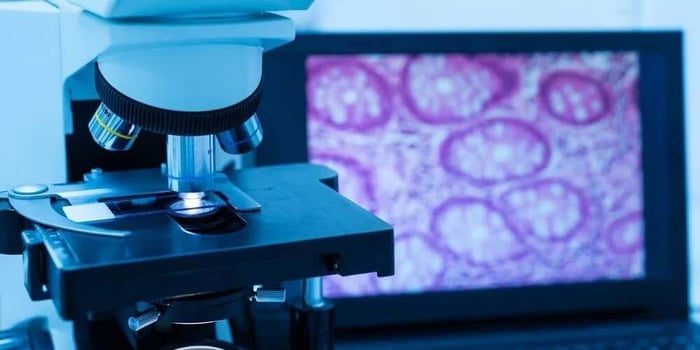
Non-psychoactive fungi have become a major focus in educational microscopy, with species like Lion’s Mane, Reishi, Shiitake, and Turkey Tail offering rich structural detail and predictable morphology for researchers. These species fall into the category of Functional Spores—spores collected from functional mushroom species and used strictly for observation, documentation, and laboratory study. As interest grows in mycology, microscopy students, hobbyists, and lab researchers are exploring how each species forms unique patterns, ornamentation, and spore characteristics that clearly differentiate one genus from another. This guide breaks down how functional mushroom spores microscopy works, what makes these non-psychoactive species so valuable for study, and how to get the most accurate slides and images from Spore Prints and suspensions.
Why Functional Mushroom Spores Microscopy Matters
Non-psychoactive species ideal for academic and lab environments
Non-psychoactive fungi such as Lion’s Mane and Reishi have become foundational in scientific and educational contexts because they allow mycologists to study spore structure legally and safely in any state. Their spores contain no psychoactive compounds, making them suitable for classrooms, university labs, and home microscopy environments.
When working with functional mushroom spores microscopy, researchers gain access to species that exhibit well-documented, stable morphological patterns. This consistency makes them reliable for comparative studies or developing microscopy skills like staining, slide preparation, and photomicrography.
Consistency and clarity in spore morphology
Functional species typically produce dense, well-formed spores with predictable characteristics, which benefits students and researchers who are learning how to differentiate genera, how spore density affects visibility, how ornamentation varies between species, and how to identify germination pores, walls, and surface textures. Observing these traits under magnification provides a strong foundation for anyone studying fungal biology or microscopy technique.
Key Functional Species Used in Microscopy
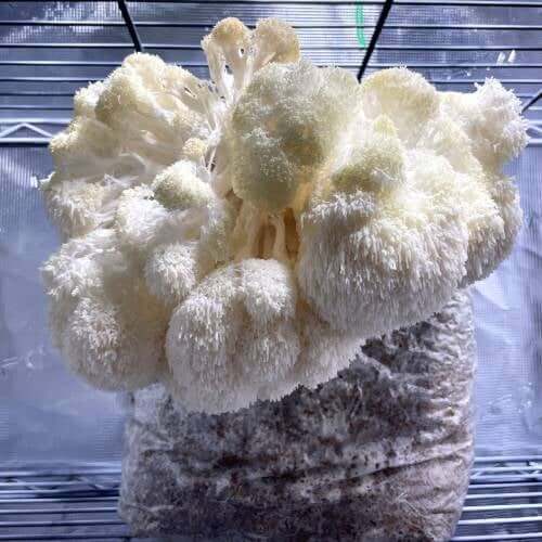
Lion’s Mane (Hericium erinaceus)
Lion’s Mane is one of the most visually distinctive spore-producing fungi in microscopy. Rather than forming gills or shelves, the organism grows cascading icicle-like formations from which it releases spores. Under magnification, Lion’s Mane spores are typically smooth, thin-walled, and ellipsoid—ideal for practicing focus, contrast adjustment, and illumination.
In functional mushroom spores microscopy, Lion’s Mane is prized for its:
Ease of visibility
Predictable shape
Uniform surface texture
Because its spores are highly consistent, they help newcomers recognize how minor structural differences appear across magnification levels.
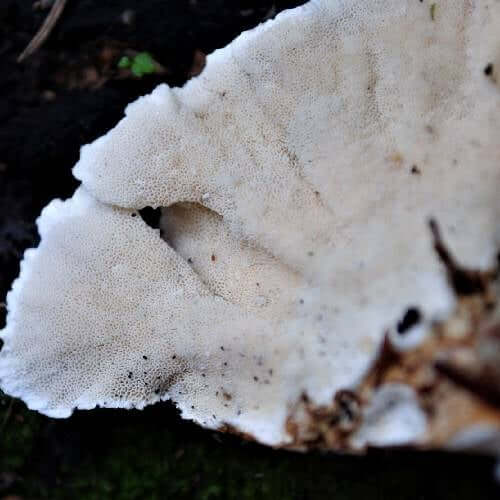
Reishi, Shiitake, Turkey Tail, and other functional genera
Beyond Lion’s Mane, many other functional species offer rich material for study.
- Reishi: Reishi spores have unique double-walled structures and ornamentation that reflect light differently from the spores of other functional species. Their color and density help students understand staining techniques and contrast adjustments.
- Shiitake: Shiitake spores are smaller and more delicate, making them ideal for practicing fine-focus adjustments and preparing thin slides.
- Turkey Tail: Turkey Tail spores showcase variation in shape and ornamentation. Their elliptical-to-cylindrical structure is useful for illustrating how different spores respond to brightfield or contrast-enhancing lighting.
Across all these species, researchers examine Functional Spores to understand how spore characteristics differ even within the same ecological or functional category.
Working With Spore Prints in the Lab
Proper handling, slide preparation, and dilution
Spore Prints remain one of the most reliable ways to begin microscopy. When handling a print, the goal is to transfer spores cleanly and avoid creating clumps that obscure fine detail.
A typical workflow includes gently scraping a small amount of material from the print, spreading a thin layer onto a clean slide, adding a drop of sterile water or microscopy mounting solution, and applying a coverslip with minimal air bubbles. For dense prints—common with functional species—a dilution may be needed to avoid overly saturated slides.
Observing patterns, ornamentation, and density
Once on the slide, spores reveal their patterns under magnification. Researchers look for wall thickness, surface texture, coloration, presence or absence of a germination pore, and clustering behavior. Functional species offer clarity in these observations, allowing easy comparison between genera.
Lion's Mane Mushroom Spores

$9.99
$19.99
Lion's Mane Mushroom Spores Lion's Mane Mushroom Spores Review Unlock the potential of your next microscopy project with our Lion's Mane Liquid Culture. This 10mL syringe is packed with a high-potency strain that’s been sterilized and sealed for your convenience.… read more
Microscopy Techniques for Functional Spores
Magnification levels and lighting choices
Different microscopy techniques highlight different aspects of Functional Spores. At 100–400x magnification, researchers examine shape and size. At 400–1000x, ornamentation and pore structures become visible. Brightfield lighting reveals outlines and density, while phase contrast highlights internal structures. Darkfield lighting emphasizes reflective ornamentation and surface textures.
Documenting and photographing spore structures
For scientific recordkeeping, it is essential to document microscopy sessions. Researchers often capture high-resolution images at multiple magnifications, note light sources and angle, record species and date, and compare new images to reference libraries. Functional mushroom spores microscopy lends itself to consistent, repeatable photography, making these species ideal for building spore comparison portfolios.
Comparing Functional Spores Across Species
Shape and ornamentation differences
Shape is one of the first characteristics researchers examine. Functional Spores vary widely: Lion’s Mane spores are smooth and ellipsoid; Reishi spores are double-walled with distinctive textures; Shiitake spores are narrowly elliptic; and Turkey Tail spores may show slight ornamentation. These differences highlight how important species selection is when building a research library.
Color, size, and density variations
Under the microscope, coloration reflects pigmentation, wall thickness, and lighting. Reishi spores may appear brownish, while Shiitake spores remain pale. Density also differs: some spores cluster naturally, while others disperse evenly, influencing how internal structures appear under magnification.
Ideal Storage and Long-Term Handling
Protecting spore material from moisture and light
To maintain the integrity of Functional Spores, proper storage is essential. Spore Prints and samples should be kept:
- In cool, dry conditions
- Away from direct sunlight
- Sealed in moisture-resistant packaging
- Labeled clearly with species and date
Moisture can cause clumping, which compromises microscopy clarity. Light can fade pigmented spores, making them more difficult to analyze.
Labeling and cataloging for research organization
A systematic approach helps researchers build reliable libraries of functional species. Many catalog Spore Prints by:
Species
Date of collection
Location of origin
Spore morphology notes
Storage method
This organization ensures the spores remain valuable for long-term study and comparison.
Final Thoughts
Functional mushroom spores microscopy is one of the most accessible and rewarding areas of mycological study. Species like Lion’s Mane, Reishi, Shiitake, and Turkey Tail provide clear, intricate spores that maintain structural integrity across slides. With the help of Spore Prints and careful technique, researchers can document, compare, and understand fungal morphology with scientific accuracy.
These non-psychoactive species establish a foundation for exploring fungal biology without legal complexities. Whether you are expanding a research library, testing new optics, or refining illumination techniques, Functional Spores offer a rich canvas for observation and growth as a microscopist.
FAQs
Are functional mushroom spores legal to observe under a microscope?
Yes. Functional species like Lion’s Mane, Reishi, Shiitake, and Turkey Tail contain no restricted compounds and are legal to study for educational microscopy.
What equipment do I need to start studying Functional Spores?
Most researchers use a compound microscope with 100–1000x magnification, slide-preparation tools, and clean Spore Prints or spore suspensions. Optional tools like phase contrast or digital cameras enhance documentation.
How long do Spore Prints remain viable for microscopy?
When stored properly—cool, dry, and protected from light—Spore Prints can remain suitable for microscopy for years, retaining their structural clarity for long-term study.