
Table of Contents
- Why Photomicrography Matters in 2025/2026
- Microscopy Gear Buying Guide (Updated for 2025/2026)
- Preparing Your Spore Slide for Imaging
- Camera Setup: Guide
- Lighting: The Secret to Clear Spore Images
- How to Photograph Mushroom Spores (Step-By-Step)
- Using Focus Stacking for Professional-Quality Images
- How to Document, Label, and Organize Your Photomicrographs
- Buying Guide: Functional Spores & Mushroom Liquid Spores for Photomicrography
- How to Edit and Prepare Images for Publication
- Final Checklist: 2025/2026 Professional Photomicrography Workflow
- Conclusion
- FAQs
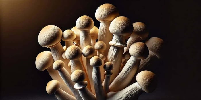
Documenting microscopy work has evolved dramatically in 2025/2026, especially for researchers, hobbyists, and mycology enthusiasts working with Functional Spores and Mushroom Liquid Spores. Whether you study Lion's Mane Mushroom Spores, carefully analyze Reishi Mushroom Spores, compare Shiitake Mushroom Spores, or capture the detailed ornamentation of Turkey Tail Mushroom Spores, clear imaging is essential for research credibility, digital archiving, and educational use.
In this complete buying guide and workflow tutorial, you’ll learn how to capture professional-grade photomicrographs, how to choose the right optical gear, how to stabilize your camera setup, and how to format your images for publications, spore documentation sheets, and microscopy portfolios. This guide is built for Magic Spore Labs customers and all microscopy users wanting clear, detailed, repeatable images.
Why Photomicrography Matters in 2025/2026
High-quality microscopy documentation serves several important purposes:
- Creates a verifiable record of mushroom spores and morphology
- Helps researchers compare Functional Spores across strains
- Supports identification work and cross-strain analysis
- Improves the scientific value of Mushroom Liquid Spores research
- Standardizes observation methods for lab-level accuracy
Clear imagery is especially important when analyzing the micro-details of Lion’s Mane Mushroom Spores or comparing the elliptical curvature of Reishi Mushroom Spores. With advances in camera sensors, LED illumination, and digital stacking software, it’s never been easier or more necessary to document your microscopy accurately.
Microscopy Gear Buying Guide (Updated for 2025/2026)
Your imaging quality begins with the right equipment. Below is a comparison of essential gear categories to help you upgrade or optimize your setup.
Microscope Comparison Table (2025/2026 Models)
| Category | Best For | Key Features | Why It Matters |
|---|---|---|---|
| Research-Grade Compound Microscope | High-resolution spore documentation | Plan-Apo objectives, Köhler illumination | Essential for detailed Functional Spores imaging |
| Digital Microscope (4K Sensor) | Fast capture + live teaching | Integrated camera, touchscreen controls | Great for Mushroom Liquid Spores and class demos |
| Phase Contrast Microscope | Transparent or colorless spores | Phase plates, halo reduction | Enhances visibility of Reishi and Turkey Tail spores |
| Portable Field Microscope | Mobile research | USB-C powered, compact objectives | Useful for in-the-field Functional Spores logging |
Choosing the right tool dramatically affects the clarity of your spore ornamentation and allows consistent documentation across multiple mushroom species.
Preparing Your Spore Slide for Imaging
This step often determines whether your final images look sharp or muddy. Follow these 2025/2026-optimized techniques for best results.
1. Use Fresh & Clean Samples
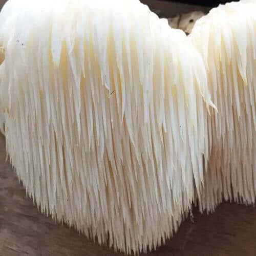
Start with high-quality Functional Spores or Mushroom Liquid Spores. Clean, unclumped spores produce sharper images and prevent shadowing. This applies equally to Lion’s Mane Mushroom Spores, Reishi Mushroom Spores, Shiitake Mushroom Spores, and Turkey Tail Mushroom Spores.
2. Avoid Overcrowding the Slide
Too many spores can overlap and block fine details. A light dispersion gives you clearer morphology and better image stacking potential.
3. Use the Correct Mounting Medium
- Distilled water for basic observation
- Lactic acid or Melzer’s for advanced ornamentation analysis
- Immersion oil for 1000x fine-detail photography
The mounting medium affects clarity, contrast, and the visible shape of spore ornamentation. For example, Shiitake Mushroom Spores often photograph best with a simple aqueous medium, while Reishi and Turkey Tail may benefit from higher contrast agents.
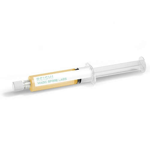
Reishi Mushroom Spores

$9.99
$19.99
Reishi Mushroom Spores Reishi Mushroom Spores Review Discover the extraordinary potential of Reishi Mushroom Spores with our ready-to-use liquid culture, designed to elevate your microscopy research. Packed in a convenient 10mL syringe, this high-potency strain is sterilized, sealed, and ready… read more
Camera Setup: Guide
Your choice of camera and mounting system can dramatically improve your photomicrography workflow.
DSLR vs Mirrorless vs Digital Microscope Cameras
| Camera Type | Strengths | Best Use |
|---|---|---|
| DSLR (APS-C) | High detail, interchangeable lenses | Publication-grade Functional Spores imaging |
| Mirrorless (4K/6K) | No vibration, superior color accuracy | Vivid Mushroom Liquid Spores visualization |
| Microscope Camera | Direct mount, simple workflow | Routine spore morphology documentation |
Essential Accessories
- Camera-to-trinocular adapter
- Remote shutter or app-trigger
- Vibration-dampening pad
- LED brightness controller
- Image stacking software
Lighting: The Secret to Clear Spore Images
Illumination is the most overlooked variable in microscopy. With the right lighting adjustments, even small spores like Turkey Tail Mushroom Spores can appear crisp and detailed.
Use Köhler Illumination
This technique ensures uniform brightness across your field of view and eliminates artifacts that often appear during high-magnification imaging.
Adjust Contrast and Condenser Height
Raising or lowering your condenser dramatically affects the contrast of translucent spores, especially for Reishi Mushroom Spores and Lion’s Mane Mushroom Spores.
How to Photograph Mushroom Spores (Step-By-Step)
This practical workflow ensures clean, repeatable, publication-ready images.
- Step 1: Clean the Optics: Use lens paper on eyepieces and objectives. Dust can mimic spore ornamentation—especially problematic in documentation work.
- Step 2: Center and Focus Your Sample: Move spores into your focal plane before attaching the camera. This reduces camera shake and keeps your exposure consistent.
- Step 3: Stabilize the Camera: Use a remote shutter or mirror-lockup (DSLR) to eliminate micro-vibration that can blur the edges of Shiitake Mushroom Spores and Turkey Tail Mushroom Spores.
- Step 4: Adjust Exposure and White Balance: Proper exposure prevents blown-out highlights and helps document accurate spore color—essential for Functional Spores work.
- Step 5: Capture Multiple Depth Layers: Even the best cameras have a limited depth of field at high magnification. Use stacking for crisp ornamentation.
Using Focus Stacking for Professional-Quality Images
Focus stacking combines multiple layers of focus to produce a single hyper-sharp image.
When to Use Stacking
- When photographing overlapping Mushroom Liquid Spores
- When documenting 3D ornamentation on Reishi Mushroom Spores
- When analyzing shape differences in Shiitake Mushroom Spores
- When capturing the fine ridges on Turkey Tail Mushroom Spores
Essential Stacking Tips
- Use small focus increments (1–3 microns)
- Capture 5–30 layers depending on magnification
- Ensure consistent lighting across shots
- Avoid moving or rotating the slide
How to Document, Label, and Organize Your Photomicrographs
Clean image organization is key for scientific, educational, or personal use. A consistent naming system helps compare Functional Spores or track the development of Mushroom Liquid Spores over time.
Recommended Naming Structure
Species_Strai n_Medium_Magnification_Date
Metadata You Should Store
- Species (ex: Lion’s Mane Mushroom Spores)
- Temperature & humidity at time of slide prep
- Magnification level
- Mounting medium
- Camera settings
- Microscope model
Over time, this creates a powerful, searchable archive for all your microscopy work.
Buying Guide: Functional Spores & Mushroom Liquid Spores for Photomicrography
High-quality photography begins with high-quality spores. Below is a comparison table including the four featured products: Lion’s Mane Mushroom Spores, Reishi Mushroom Spores, Shiitake Mushroom Spores, and Turkey Tail Mushroom Spores.
Functional Spore Comparison Table
| Spore Type | Shape & Size | Best Medium | Best For |
|---|---|---|---|
| Lion's Mane Mushroom Spores | Elliptical, smooth | Distilled water | Beginner spore imaging, morphology teaching |
| Reishi Mushroom Spores | Brownish, double-walled | Lactic acid | Advanced morphology & structural imaging |
| Shiitake Mushroom Spores | Elliptical, hyaline | Water or IPA | General microscopy & contrast tests |
| Turkey Tail Mushroom Spores | Small, transparent | Phase contrast | High-detail imaging & stacking practice |
All four of these products, when prepared carefully, produce publication-quality microscopy images ideal for a documentation workflow.
How to Edit and Prepare Images for Publication
Editing should enhance clarity while avoiding manipulation that alters the biology.
Standard Editing Workflow
- Crop to remove blank slide edges
- Adjust levels to improve contrast
- Increase sharpness slightly (10–20%)
- Use noise reduction sparingly
- Maintain natural color
Your goal is accuracy, not exaggeration.
Final Checklist: 2025/2026 Professional Photomicrography Workflow
- Use high-quality Functional Spores or Mushroom Liquid Spores
- Prepare slides with clean, unclumped samples
- Use Köhler illumination for even lighting
- Stabilize your camera to prevent micro-blur
- Capture multiple focal layers for stacking
- Label and store metadata consistently
- Export clean, accurate, publication-ready images
When following these steps, your Lion’s Mane Mushroom Spores, Reishi Mushroom Spores, Shiitake Mushroom Spores, and Turkey Tail Mushroom Spores images will showcase the clarity, detail, and precision expected in 2025/2026 microscopy documentation.
Conclusion
Photomicrography is no longer just a technical skill—it’s becoming a core requirement for researchers, educators, and microscopy enthusiasts working with Functional Spores and Mushroom Liquid Spores. With improved camera technology, advanced microscope optics, and powerful stacking tools, your ability to capture professional-grade spore images has never been greater.
By choosing quality spore samples like Lion’s Mane Mushroom Spores, Reishi Mushroom Spores, Shiitake Mushroom Spores, and Turkey Tail Mushroom Spores, and by following this modernized 2025/2026 workflow, you can create clear, consistent, publishable microscopy documentation that elevates your research and educational materials.
FAQs
What magnification do I need to properly photograph mushroom spores?
Most mushroom spores are best photographed between 400x–1000x, with 1000x providing the clearest detail for morphology and ornamentation.
Do I need a dedicated microscope camera, or can I use my phone or DSLR?
You can use any of these, but a dedicated microscope camera offers the most consistent, publication-ready imaging workflow.
How do I know if my spore images are documented correctly for research use?
Your image is properly documented when it has clean lighting, accurate color, sharp morphology, and complete metadata for repeatable observations.