

Table of Contents
- Why Contaminants Matter in Mushroom Microscopy
- Quick Reference Table: Contaminants vs. True Fungal Spores
- The 10 Most Common Microscopy Contaminants Explained
- Buying Guide: Best Tools for Clean Microscopy Slides in 2025
- What Real Mushroom Spores Should Look Like
- Step-by-Step: How to Prepare a Clean Microscopy Slide
- Troubleshooting: Is It a Contaminant?
- Conclusion
- FAQs
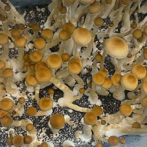
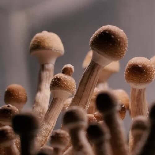
Contamination is one of the most common challenges for both new and experienced microscopists. Whether you're examining Mushroom Liquid Spores, studying Spore Prints, or documenting slides for research, contaminants can distort results, mimic fungal structures, and complicate observation. This guide gives you a clear, practical, and image-centered explanation of what the most common contaminants look like, why they appear, and exactly how to avoid them in your microscopy setup. It is written as a buying guide, using direct comparisons and step-by-step instructions to help you produce cleaner, more accurate microscopy slides in 2025 and beyond.
Why Contaminants Matter in Mushroom Microscopy
Clean sample preparation is the foundation of accurate microscopy. When working with Mushroom Liquid Spores or traditional Spore Prints, anything that enters your slide—dust, lint, bacteria, pollen, fibers, or moisture droplets can alter what you see through the eyepiece. Many contaminants even resemble fungal spores, hyphae, or cystidia, creating confusion for beginners who are still learning how to differentiate microscopic structures.
Beyond simple visual interference, contaminants can lead to incorrect species identification, misinterpreted morphology, or misleading documentation. As microscopy grows more popular in 2025, so does the need for updated contamination-prevention techniques, especially when using sensitive high-magnification objectives.
Quick Reference Table: Contaminants vs. True Fungal Spores
| Microscopy Element | Appearance Under 400–1000x | Common Confusion | Key Identifier |
|---|---|---|---|
| Dust Particles | Irregular, jagged, inconsistent shapes | Spores with deformities | Lack of uniform structure |
| Pollen Grains | Spiky, patterned spheres | Thick-walled spores | Surface ornamentation |
| Textile Fibers | Long strands, often frayed | Hyphae or mycelial fragments | No septa, bends sharply |
| Yeast Cells | Small budding ovals | Tiny spores | Budding patterns |
| Airborne Mold Spores | Dark, round clusters | Fungal spore groups | Mixed sizes and clumping |
Penis Envy Mushroom Spore Syringe

$22.99
$24.99
PE#6 (Penis Envy 6) Mushroom Spore The Penis Envy #6 Mushroom Spore Syringe from Magic Spore Labs delivers basidiospores from the most systematically refined expression of the Penis Envy lineage available in the Psilocybe cubensis research catalog — a sixth-generation… read more
The 10 Most Common Microscopy Contaminants Explained
1. Household Dust
Dust is the most widespread contaminant found when preparing slides. It enters the field of view during sample transfer, mounting fluid application, or cover slip placement. Under magnification, dust appears as jagged, amorphous debris with no predictable geometry. When working with Mushroom Liquid Spores or rehydrated Spore Prints, dust can disguise itself as malformed spores.
To prevent dust contamination, clean your workspace, keep slides covered until use, and use lint-free wipes before mounting samples.
2. Textile Fibers and Lint
Fibers from clothing, lab coats, wipes, or paper towels are extremely common contaminants. They show up as long, threadlike strands that bend irregularly and often display frayed edges. Their shape makes them easy to confuse with hyphae, but unlike true hyphae, textile fibers lack septa and typically vary widely in thickness.
3. Airborne Spores From Other Fungi
When working around multiple Spore Prints or preparing several microscopy slides in one session, airborne spores from unrelated fungi can drift into your sample. These contaminants often appear as unfamiliar spore shapes, sometimes smaller or rounder than the Mushroom Liquid Spores you're examining.
4. Pollen Grains
Pollen is one of the easiest contaminants to accidentally introduce. It is highly airborne and can enter through subtle airflow, HVAC movement, or seasonal exposure. Under the microscope, pollen looks highly decorative—spiky, patterned, often bright. Because of their size and ornamentation, they might be confused with exotic fungal spores.
5. Yeast Cells
Yeast contaminants appear as tiny oval structures that frequently show budding patterns. When rehydrating Spore Prints or preparing Mushroom Liquid Spores for observation, yeast can be introduced through non-sterile water or unclean tools. Yeast typically forms clusters or chains that reveal their biological patterning.
6. Bacteria (Rod and Cocci)
Bacteria are smaller than most fungal spores, but at high magnification (1000x oil), they become more visible. These contaminants appear as rod-shaped or circular structures arranged in lines, clusters, or chains. They may appear when using tap water or improperly stored slide preparation tools.
7. Mold Hyphae
Mold hyphae resemble fungal hyphal structures but often come from airborne molds unrelated to the species you're examining. Unlike textile fibers, these hyphae may show branching and uniform width. They contaminate samples stored in humid environments or left uncapped for too long.
8. Insect Scales and Hair
Insect scales look like tiny feathered shapes with textured surfaces. Human or animal hair also appears as large, tube-like strands. Both can drift onto microscopy slides if samples are prepared near carpets or high-traffic areas.
9. Residual Agar, Substrate, or Media Particles
When transferring spores, substrate or agar fragments may become mixed in with the sample. These appear as opaque, irregular structures that lack the smooth walls of true spores. This is especially seen when preparing Mushroom Liquid Spores from a culture medium.
10. Moisture Bubbles and Oil Droplets
Too much mounting fluid or improper cover slip pressure can trap air bubbles. These bubbles mimic round structures, confusing beginners who mistake them for spherical spores or cystidia. Proper droplet size and angled cover slip placement solve this issue.
Buying Guide: Best Tools for Clean Microscopy Slides in 2025
This section highlights the tools and supplies that deliver the cleanest microscopy experience when working with Spore Prints or Mushroom Liquid Spores. These items help reduce contamination, increase clarity, and improve your imaging workflow.
| Equipment | Best Use | Why It Helps | What to Look For |
|---|---|---|---|
| Lint-Free Wipes | Cleaning slides | Reduces dust and fibers | Anti-static, lab-grade materials |
| Pre-Sterilized Water | Rehydrating Spore Prints | Prevents yeast and bacteria | Hermetically sealed vials |
| Disposable Pipettes | Mount droplet control | Consistent droplet size | Fine-tip micro droplet control |
| Slide Storage Boxes | Safe transport | Limits airborne contamination | Dustproof silicone-lined cases |
| Still-Air Box | Advanced prep | Reduces airflow contamination | Clear walls, comfortable hand ports |
What Real Mushroom Spores Should Look Like
To recognize contaminants, you must understand how real spores appear under the microscope. When examining Mushroom Liquid Spores or freshly hydrated Spore Prints, the following characteristics should be visible:
- Consistent elliptical or subelliptical shapes
- Defined, thick-walled structures
- Uniform spore size within the same species
- Germ pores appearing as lighter zones at one end
- Smooth, matte, or occasionally textured walls depending on species
Magic Spore Labs offers clean, high-clarity microscopy materials designed to minimize contaminants before your slide is even prepared.
Step-by-Step: How to Prepare a Clean Microscopy Slide
Use this contamination-free workflow whenever preparing Mushroom Liquid Spores or a rehydrated Spore Print sample:
- Prepare Your Workspace: Wipe surfaces with alcohol, reduce airflow, and keep all tools within reach.
- Clean Your Slides: Use lint-free wipes or sterile slide packs to avoid fingerprints and fibers.
- Hydrate the Sample: Use sterile water to hydrate Spore Prints or evenly mix Mushroom Liquid Spores.
- Use a Controlled Mounting Droplet: A tiny droplet—small enough not to overflow—is ideal. Excess liquid increases bubble formation.
- Angle the Cover Slip: Lower the cover slip from one edge to reduce bubble formation and trap fewer airborne particles.
- Seal or Store: For long-term observation, seal the slide edges. Otherwise, store slides in a dustproof case.
Troubleshooting: Is It a Contaminant?
| What You See | Likely Contaminant | Common Confusion | Quick Fix |
|---|---|---|---|
| Bubbles or rings | Moisture artifacts | Rounded spores | Use smaller droplets |
| Long fibers | Lint or textile threads | Hyphae | Use lint-free wipes |
| Large spiky circles | Pollen | Decorated spores | Limit airflow |
| Dark clusters | Mold spores | Fungal spore groups | Store samples sealed |
Conclusion
Microscopy is an incredibly rewarding field, but the clarity of your results depends on mastering contamination control. Whether you're examining Mushroom Liquid Spores or studying Spore Prints from rare species, the techniques and tools outlined in this guide will dramatically improve the accuracy of your imaging. Cleaner slides mean clearer images, more consistent documentation, and more reliable microscopy observations overall.
FAQs
Why do contaminants show up when observing Mushroom Liquid Spores?
Contaminants often enter the slide during preparation, especially from dust, fibers, or unsterile water. Because Mushroom Liquid Spores are hydrated and fluid-based, particles can easily stick to the sample if the workspace or tools are not cleaned properly.
How can I tell the difference between contaminants and real spores from Spore Prints?
True spores from Spore Prints will show uniform shapes, defined walls, and consistent size ranges. Contaminants like lint, pollen, or dust appear irregular, textured, or unusually large. Consistency is the biggest indicator of real spores.
What tools help prevent contamination when preparing microscopy slides?
Using lint-free wipes, sterile water, clean slides, disposable pipettes, and dustproof cases significantly reduces contamination. These tools help keep samples of Mushroom Liquid Spores or Spore Prints clean throughout the preparation process.