
Table of Contents
If you’re spending time behind a microscope, Gulf Coast Spores are a fascinating sample to document. Known for their resilience and consistency under observation, these spores are often chosen by microscopy enthusiasts who want clear structures, reliable visibility, and repeatable results. This guide is written specifically for lab-style observation and documentation, focusing on what you can see, how to tell real features from artifacts, and how to handle samples responsibly.
Everything here is designed to help you slow down, observe carefully, and take useful notes—without overcomplicating the process or drifting into speculation. Whether you’re newer to microscopy or refining your technique, this checklist-style approach keeps things practical and repeatable.
High-Level Overview
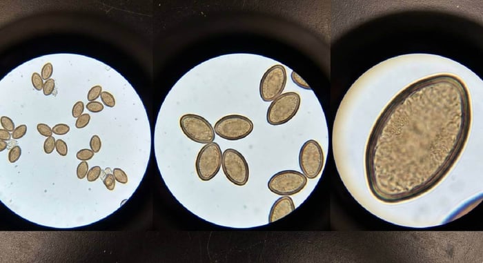
Under a microscope, Gulf Coast spores typically present a balanced combination of clarity and density. This makes them useful for both casual observation and more detailed documentation sessions. When prepared properly, the sample often reveals well-defined spore outlines that are easy to track across magnification levels.
What makes these spores especially approachable is their tendency to show consistent visual traits across slides. That consistency allows observers to focus less on troubleshooting and more on pattern recognition, measurement practice, and comparison between slides.
At lower magnifications, you’ll usually notice clusters forming loose groupings rather than tight stacks. As you increase magnification, individual spores become easier to isolate visually, helping you practice edge definition and contrast control.
Many microscopy hobbyists appreciate Gulf Coast spores because they offer:
- Clear visibility without excessive debris
- Reliable shape consistency from slide to slide
- Good contrast response with basic lighting adjustments
- Predictable appearance that supports repeat documentation
This reliability is ideal for note-taking, sketching, and learning how small changes in focus or lighting affect what you see.
Observation Checklist
Approaching microscopy with a checklist mindset helps prevent rushed conclusions and missed details. Use the following points as a guide while observing Gulf Coast spores.
Spore Shape
Begin by identifying the general outline. Most observers note an oval to slightly elongated form. Focus on whether the edges appear smooth or irregular, and whether the shape remains consistent across multiple spores within the same field of view.
Ask yourself:
- Are the spores uniform in outline?
- Do any appear collapsed or distorted?
- Is shape consistency maintained when you refocus?
Wall Definition
Spore walls are one of the easiest features to misinterpret. With Gulf Coast mushroom spores, wall boundaries are usually visible without extreme adjustments. Slowly adjust focus to see whether the wall appears as a clean edge or fades in and out.
A clearly defined wall typically stays visible through minor focus changes, while artifacts often disappear or shift dramatically.
Color and Transparency
Under standard brightfield lighting, these spores often appear translucent with subtle coloration. Avoid over-adjusting brightness, as washed-out lighting can mask fine detail.
Look for:
- Even coloration across the spore body
- Subtle internal shading rather than solid darkness
- Consistency between spores in the same sample
Internal Features
Some observers report faint internal variations or gradients. These should appear stable as you adjust focus. If an internal “feature” moves independently or disappears with slight lighting changes, it’s more likely an optical artifact.
Patience is key here—spend time observing the same spore rather than constantly moving the slide.
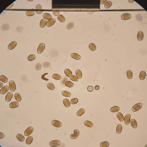
Distribution and Density
Scan multiple areas of the slide. Gulf Coast spores are often distributed evenly, making it easier to compare regions. Uneven clustering may indicate slide preparation issues rather than natural variation.
Document density differences between fields of view to build a more complete record.
Gulf Coast Mushroom Spore Syringe

$24.99
Gulf Coast Spores Discover the remarkable Gulf Coast Liquid Spores, a favorite among spore researchers worldwide for its renowned Gulf Coast strain of Psilocybe cubensis. Originally discovered along the Gulf Coast's humid and warm regions, this strain has become a… read more
Common Artifacts vs True Features
One of the biggest challenges in microscopy is learning what not to record. Artifacts can be convincing, especially to the untrained eye. Understanding common pitfalls helps keep your notes accurate.
Air Bubbles
Air bubbles are among the most common distractions. They often appear perfectly round with a thick, dark outline. Unlike spores, bubbles may show strong light refraction and can shift slightly if the slide is tapped.
True spores maintain shape and position even with minor slide movement.
Dust and Debris
Dust particles can appear irregular and sharply opaque. They often lack consistent shape and may cast uneven shadows. Unlike Gulf Coast spores, debris rarely shows uniformity across the slide.
If a structure looks dramatically different from surrounding elements, it’s worth questioning.
Optical Illusions
Changing focus can create the illusion of internal structures or halos. These effects usually appear and disappear rapidly as you adjust the fine focus knob.
A true feature remains visible across a narrow focus range, while an illusion flickers or vanishes entirely.
Slide Preparation Marks
Scratches or residue on slides can mimic linear features. Rotate the slide slightly—true spore features rotate with the sample, while slide imperfections remain fixed relative to the lens.

Storage & Handling
Proper storage and handling preserve sample integrity and make future observation more consistent. Even for microscopy-only purposes, small habits can have a big impact.
Temperature Awareness
Store samples in a stable, cool environment away from direct heat sources. Extreme temperature changes can affect fluid clarity and introduce condensation, which complicates observation.
Light Exposure
Prolonged exposure to strong light can degrade visibility over time. Keep samples in opaque containers or drawers when not in use to maintain consistency between sessions.
Clean Tools Matter
Always use clean slides, cover slips, and droppers. Residue from previous sessions increases the chance of confusing artifacts. A quick cleaning routine saves time later.
Labeling and Notes
Label slides clearly with dates and sample identifiers. Pair visual observations with written notes describing magnification, lighting conditions, and focus behavior. Over time, these notes become invaluable references.
Conclusion
Observing Gulf Coast Spores under a microscope is as much about patience as it is about technique. Their consistent appearance makes them a rewarding subject for careful documentation, comparison, and learning. By using a structured checklist, understanding common artifacts, and handling samples responsibly, you can build reliable observation habits that improve with every session.
Microscopy isn’t about rushing to conclusions it’s about noticing small details, questioning what you see, and keeping clear records. With the right approach, each slide becomes an opportunity to refine your skills and deepen your understanding of what’s happening under the lens.
FAQs
Are Gulf Coast spores easy to observe for beginners?
Many microscopy users find them approachable due to their consistent appearance and clear outlines. This makes them suitable for learning how to adjust focus, lighting, and magnification.
Why do some spores look different on the same slide?
Variation can come from preparation differences, focal plane changes, or overlapping material. Observing multiple areas helps determine whether differences are meaningful or incidental.
How long can samples remain usable for observation?
When stored properly, samples can remain visually stable for extended periods. Consistent storage conditions help maintain clarity across sessions.
What magnification works best?
Many observers start low to locate areas of interest, then increase magnification gradually. This layered approach reduces eye strain and improves feature recognition.