

Table of Contents
Cordyceps spores sit in a fascinating corner of mycology. They don’t behave, look, or even originate the same way as many familiar mushroom spores, and that difference is exactly why they draw so much attention in research settings. Whether you’re cataloging specimens, building a microscopy reference library, or simply expanding your understanding of less conventional fungi, Cordyceps spores offer a distinctly different experience from the start.
This guide takes a research-first look at Cordyceps spores what Cordyceps is at a high level, why it’s studied so closely, how to document spores under the microscope, and the most common questions researchers tend to ask when encountering them for the first time.
What Cordyceps Is (High-Level)
Cordyceps refers to a broad genus of fungi rather than a single mushroom type. Unlike the classic cap-and-stem mushrooms most people picture, many Cordyceps species are known for their parasitic or symbiotic relationships with insects and arthropods. This ecological niche alone sets them apart from wood-loving or soil-based fungi.
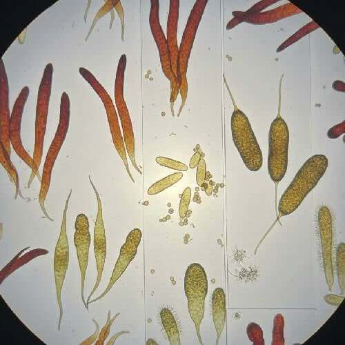
From a structural standpoint, Cordyceps species often produce elongated, club-like fruiting bodies instead of umbrella-shaped caps. These fruiting structures are designed to release spores in environments where precision matters more than volume. As a result, Cordyceps spores tend to display unique shapes, surface textures, and dispersal characteristics.
Another key distinction is diversity. The Cordyceps genus includes hundreds of species, each adapted to a specific host or environment. That diversity means spores can vary widely in size, segmentation, pigmentation, and behavior when mounted on slides. For microscopy-focused researchers, this makes Cordyceps a broad and rewarding category to explore rather than a single, uniform subject.
Why Cordyceps Is Studied
Cordyceps attracts attention across multiple research disciplines, and spores are often the starting point for deeper study. Mycologists, microscopists, and taxonomists are drawn to Cordyceps because it challenges conventional fungal assumptions.
One reason Cordyceps mushroom spores are studied is classification. Many species can look similar at the macroscopic level, but their spores reveal subtle distinctions that help differentiate one species from another. Spore shape, septation, and ornamentation often hold the clues needed for accurate identification.
Another reason is lifecycle analysis. Cordyceps species tend to have complex reproductive cycles tied closely to their hosts. Studying spores under the microscope allows researchers to document how these fungi propagate, adapt, and persist in highly specialized environments.
Cordyceps is also studied because it bridges multiple ecological roles. It intersects mycology, entomology, and environmental science in a way few fungi do. Spores represent the transferable, observable link between these systems, making them a valuable subject for cross-disciplinary research.
Finally, Cordyceps mushroom spores are studied simply because they behave differently. They don’t always disperse or germinate in predictable ways, and that unpredictability pushes researchers to refine observation techniques, documentation standards, and classification frameworks.
Cordyceps Mushroom Spores

$9.99
$19.99
Cordyceps Mushroom Spores Cordyceps Mushroom Spores Review Unlock the potential of your mycological studies with our premium Cordyceps Mushroom Spores. This 10mL syringe contains a high-potency strain in a ready-to-use liquid culture, ensuring you have everything you need for successful… read more
Microscopy Documentation Checklist
Because Cordyceps mushroom spores vary so widely, consistent documentation is critical. A structured approach helps ensure observations are comparable across sessions, specimens, and research logs.
- Spore Shape and Structure: Begin by noting the overall form. Cordyceps mushroom spores are often elongated or filament-like, sometimes appearing segmented or divided into multiple cells. Record whether the spore appears straight, curved, or irregular.
- Size Measurements: Measure length and width using a calibrated eyepiece or imaging software. Even small size differences can be significant when comparing closely related species.
- Septation and Segmentation: Many Cordyceps mushroom spores show internal divisions. Document the number of segments and how clearly defined they are. This detail is frequently used in species differentiation.
- Surface Texture: Under higher magnification, look for smooth, rough, or ornamented surfaces. Some spores may appear glassy, while others show faint ridges or irregularities.
- Color and Transparency: Note whether spores are clear, lightly pigmented, or darker in tone. Changes in color under different lighting conditions are also worth recording.
- Mounting Medium Used: Always log whether spores were observed in water, glycerin, or another medium. Cordyceps spores can appear slightly different depending on how they are mounted.
- Imaging and Metadata: Capture images at multiple magnifications and label them clearly. Include date, sample source, magnification level, and any notable environmental context associated with the specimen.
This checklist doesn’t just support accuracy it builds a reference archive that becomes more valuable over time as patterns emerge across samples.
Common Questions
Are Cordyceps spores harder to identify than other spores?
They can be. The diversity within the genus means spores don’t follow a single “standard” appearance. Careful measurement and comparison are essential.
Do all Cordyceps spores look similar?
No. Spore morphology can vary dramatically between species. This variation is part of what makes Cordyceps such an interesting research subject.
Why do Cordyceps spores often appear segmented?
Segmentation is tied to how these fungi reproduce and adapt to their environments. It’s a defining feature in many species and an important classification marker.
Is special equipment required to study Cordyceps spores?
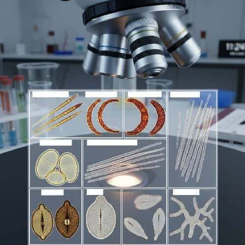
Standard compound microscopes are sufficient for most observations, but higher magnification and good lighting control make detailed documentation easier.
How should Cordyceps spores be stored for research?
Proper labeling, dry storage, and protection from light and humidity help preserve spore samples for long-term study.
Conclusion
Cordyceps spores stand apart because Cordyceps itself stands apart. Their shapes, structures, and behaviors reflect a genus that evolved along a very different path from most familiar mushrooms. For researchers, this difference isn’t a hurdle it’s the appeal.
By approaching Cordyceps mushroom spores with careful observation, consistent documentation, and an open mind, researchers gain insight into one of the most unconventional fungal groups studied today. Whether you’re building a personal microscopy archive or contributing to broader classification efforts, Cordyceps spores reward patience, precision, and curiosity.
FAQs
What makes Cordyceps spores unique compared to other fungal spores?
Their morphology, segmentation, and ecological context differ significantly from many common mushroom spores.
Can Cordyceps spores be used for comparative research?
Yes. They are often compared across species to study variation, classification, and evolutionary patterns.
Do Cordyceps spores require special handling?
They benefit from careful mounting and thorough labeling, but no unusual handling is required beyond standard microscopy practices.
Why are Cordyceps spores popular in microscopy-focused research?
Their diversity and unusual structures make them ideal for detailed observation and long-term study.