

Table of Contents
Tidal Wave Spores have gained attention among microscopy-focused collectors and researchers for their distinct lineage, visual characteristics, and consistency under magnification. While the name often sparks curiosity, the real interest lies in how these spores present themselves during observation sessions and how reliably they can be documented over time. This guide breaks down what makes Tidal Wave Spores notable from a research and microscopy perspective, keeping the focus on identification, documentation, and comparative analysis rather than cultivation.
Whether you’re building a reference library, refining your microscopy workflow, or simply expanding your understanding of spore morphology, this overview provides a clear, structured look at what to expect and how to work with Tidal Wave Spores in a responsible, research-only context.
Background & Naming
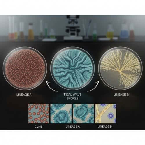
The name “Tidal Wave” reflects a hybrid origin that combines traits from two well-known genetic lines, resulting in a spore profile that many researchers describe as visually striking and relatively consistent. From a naming standpoint, it follows a common convention within spore research communities: using evocative terminology to signal a blend of characteristics rather than a single, isolated lineage.
At a high level, Tidal Wave Spores are recognized for:
- A reputation for clear, well-defined spore structures under standard microscopy ranges
- Balanced morphology that makes them approachable for both newer and experienced observers
- Consistent visual traits across reputable lab-prepared samples
It’s important to note that naming conventions are not scientific classifications. Instead, they act as shorthand within research and collector circles to describe expected visual tendencies. This makes proper documentation especially important, as individual samples can still vary based on preparation, storage, and handling.
Microscopy Documentation Checklist
Accurate documentation is the foundation of meaningful spore research. When working with Tidal Wave Spores, having a repeatable checklist helps ensure that observations are consistent and comparable over time. Below is a practical documentation framework designed specifically for microscopy sessions.
Pre-Observation Setup
- Clean slides and coverslips to avoid debris interference
- Calibrated microscope with noted magnification levels
- Consistent lighting setup (LED intensity, condenser position)
- Notebook or digital log prepared before viewing
Key Visual Features to Record
During observation, focus on capturing descriptive details rather than interpretations. Common points of documentation include:
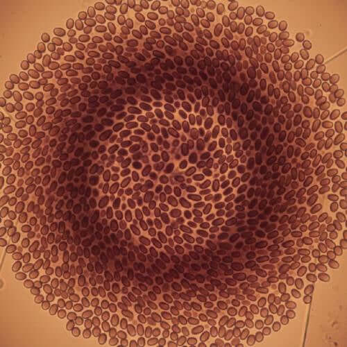
- Spore shape (elliptical, sub-elliptical, or variations)
- Wall thickness appearance
- Color tone under transmitted light
- Surface texture impressions at higher magnifications
Measurement & Imaging Notes
- Estimated size range using calibrated scale
- Magnification level used for each image or note
- Any optical adjustments made during viewing
Keeping this checklist consistent helps reduce subjective drift between sessions and ensures that observations of Tidal Wave Spores remain useful for long-term reference.
How to Compare Observations Across Sessions
One of the most valuable aspects of spore microscopy is the ability to compare observations over time. With Tidal Wave Mushroom Spores, this process becomes especially informative due to their relatively stable visual traits when properly handled.
Standardizing Your Conditions
Before comparing notes, confirm that each session followed similar conditions. Even small differences in lighting or slide preparation can alter perceived characteristics. Key factors to standardize include:
- Magnification ranges used
- Light source intensity and color temperature
- Slide mounting technique
Using Reference Frames
Establish a baseline observation from an initial session and treat it as a reference frame. When reviewing later sessions, compare new observations directly against this baseline rather than relying on memory alone.
Tidal Wave Mushroom Spore Syringe

$24.99
Tidal Wave Liquid Spores Discover the potential of Tidal Wave Liquid Spores, a premium option for mycology enthusiasts looking to explore unique mushroom varieties. These liquid spores are designed for easy inoculation, offering a smooth, hassle-free experience for both beginners… read more
Identifying Meaningful Variations
Not all differences indicate a change in the sample itself. When comparing Tidal Wave Spores across sessions, consider:
- Are variations consistent across multiple fields of view?
- Do differences align with known optical adjustments?
- Could storage time or slide age play a role?
By asking these questions, researchers can separate meaningful observations from normal microscopy variability.
Pairing Suggestions
In a research and collector setting, pairing spore samples is less about combination and more about comparative study. Tidal Waves are often included in curated bundles because they offer a useful contrast against other popular microscopy subjects.
Why Pair Tidal Wave Spores?
- They provide a balanced reference point for morphology comparison
- Their visual clarity makes them suitable for side-by-side study
- They help highlight subtle differences between lineages
Effective Comparative Pairings
When included in a bundle or study set, Tidal Waves are commonly paired with samples that:
- Exhibit more elongated or rounded spore shapes
- Show stronger color variation under similar lighting
- Have different wall thickness characteristics
This approach allows researchers to refine observational skills and build a more nuanced understanding of spore diversity without shifting focus away from microscopy.
Conclusion
Tidal Wave Spores occupy a notable place in microscopy-focused research and collecting due to their recognizable visual traits and usefulness in comparative study. By approaching them with a structured documentation process, standardized observation conditions, and thoughtful comparison methods, researchers can gain meaningful insights without relying on assumptions or informal impressions.
For anyone building a microscopy reference library, Tidal Waves offer a reliable subject that rewards careful observation and disciplined note-taking. When treated strictly as a research specimen and documented responsibly, they serve as a valuable addition to an
FAQs
What makes Tidal Wave Spores stand out under a microscope?
They are often noted for their balanced shape and consistent appearance, which can make documentation and comparison more straightforward than with highly variable samples.
Are Tidal Wave Spores suitable for beginners in microscopy?
Many researchers consider them approachable due to their clarity and predictability, though proper documentation practices are still essential regardless of experience level.
How should observations be stored for long-term reference?
Detailed written logs combined with labeled images and consistent terminology provide the most reliable long-term reference system.
Can different batches look different?
Yes. While Tidal Wave Spores are known for consistency, variations can occur based on preparation, age, and handling, which is why comparative documentation is important.