

Table of Contents
- What Is the Enigma Mutation?
- Why Enigma Mushroom Spores Are Rare
- Microscopic Characteristics of Enigma Mushroom Spores
- Recommended Equipment for Observing Mushroom Spores
- Preparing a Slide for Microscopy Observation
- Enigma vs. Standard Psilocybe Cubensis Spores
- Documentation Best Practices for Rare Specimens
- Storage and Preservation Tips
- Ethical and Legal Considerations
- Conclusion
- FAQs
The world of mushroom microscopy is filled with fascinating strains, but few generate as much curiosity as Enigma Mushroom Spores. Known for their unusual mutation and rarity within the Psilocybe cubensis lineage, Enigma has become a topic of serious interest among collectors and microscopy researchers.
This guide explores what makes this mutation unique, how it differs from traditional cubensis varieties, and how to properly observe and document Mushroom Spores in a lab setting. Whether you are building a research collection or refining your microscopy techniques, this complete breakdown will help you understand the distinct characteristics and handling considerations associated with this rare specimen.
What Is the Enigma Mutation?
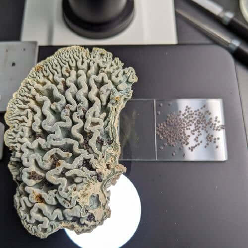
Enigma is widely recognized as a stabilized mutation of Psilocybe cubensis. Unlike typical cubensis varieties that form recognizable caps and stems, Enigma displays dense, brain-like or coral-style formations. This unusual growth pattern sets it apart visually and genetically within the cubensis family.
From a microscopy perspective, the mutation is intriguing because it often exhibits irregular or limited sporulation. In standard cubensis varieties, spore production is abundant and predictable. Enigma, however, is known for producing fewer spores, which contributes to its rarity and collector demand.
Researchers often compare it to other cubensis isolates to better understand mutation stability and lineage behavior. While the outward fruiting structure differs dramatically, microscopic analysis shows that the spores themselves share many baseline cubensis traits.
Why Enigma Mushroom Spores Are Rare
One of the defining features of Enigma Mushroom Spores is their limited availability. Because the mutation does not consistently release spores in the same volume as traditional cubensis strains, viable spore samples are less common.
This rarity impacts both collectors and researchers. Most cubensis varieties can be obtained through prints or spore syringes without difficulty. Enigma specimens, however, are often stabilized and maintained through isolated cultures rather than relying solely on heavy sporulation.
For microscopy enthusiasts, this means that acquiring verified samples requires careful sourcing and documentation. The scarcity also makes accurate labeling and storage even more important, as replacement samples are not always easily accessible.
Microscopic Characteristics of Enigma Mushroom Spores
Under magnification, Enigma Mushroom Spores generally resemble other Psilocybe cubensis spores. They are typically ellipsoid in shape with smooth outer walls and a visible germ pore. Pigmentation appears purple-brown when observed under brightfield microscopy.
At 400x magnification, researchers can clearly observe overall spore shape and color. At 1000x using oil immersion, the germ pore becomes more defined, allowing for more detailed documentation. Spore measurements typically fall within the expected cubensis range in microns, reinforcing their taxonomic classification within the species.
While the mutation alters macroscopic development, microscopic morphology remains consistent with cubensis traits. This contrast between external mutation and internal consistency makes Enigma particularly interesting for comparative study.
Recommended Equipment for Observing Mushroom Spores
Proper equipment is essential for accurate microscopy. To examine Enigma Mushroom Spores effectively, researchers should use:
- A compound microscope capable of 400x–1000x magnification
- Oil immersion lens for high-detail analysis
- Köhler illumination for improved contrast
- Clean glass slides and cover slips
- Sterile distilled water or appropriate mounting medium
- Digital camera attachment for documentation
Maintaining a stable workspace with minimal airflow helps prevent contamination and preserves sample integrity. Even though spores are examined strictly for research and identification purposes, handling standards should remain consistent with professional lab practice.
Enigma Mushroom Spore Syringe

$22.99
Enigma Spores The Enigma Mushroom Spore Syringe stands out as one of the most intriguing offerings in modern mycology, captivating researchers with its rare and unconventional genetic profile. Unlike traditional Psilocybe Cubensis varieties, Enigma is renowned for its distinct mutation… read more
Preparing a Slide for Microscopy Observation
Slide preparation plays a critical role in obtaining clear, measurable results. Begin by ensuring all surfaces and tools are clean. Place a small sample of spores onto a glass slide and apply a drop of sterile distilled water or mounting medium.
Carefully lower a cover slip to minimize air bubbles. Start viewing under low magnification to center the specimen, then gradually increase to higher magnification levels. Adjust illumination and fine focus controls to sharpen the spore wall and germ pore visibility.
Photographing and measuring spores during observation allows you to build a reliable research archive. Label each slide with date, sample identification, and magnification level used. Proper documentation strengthens comparative analysis over time.
Enigma vs. Standard Psilocybe Cubensis Spores
Although Enigma Mushroom Spores belong to the cubensis species, availability and consistency differ from common strains such as Golden Teacher or B+.
Similarities:
- Ellipsoid shape
- Smooth outer wall
- Visible germ pore
- Purple-brown coloration
Differences:
- Reduced sporulation frequency
- Less predictable spore density in prints
- Higher rarity in verified samples
For researchers conducting side-by-side comparisons, these distinctions highlight how macroscopic mutations do not always drastically alter microscopic morphology.
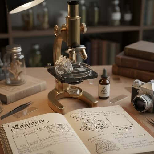
Documentation Best Practices for Rare Specimens
When working with rare Mushroom Spores, detailed documentation is essential. Keeping structured records ensures long-term research value and accurate comparisons.
Each observation session should include:
- Date of examination
- Magnification level used
- Measured spore dimensions
- Notes on germ pore visibility
- Lighting technique applied
- Photographic references
Building a dedicated microscopy journal helps track subtle variations and supports ongoing research. Rare mutations like Enigma benefit greatly from consistent measurement and archival practices.
Storage and Preservation Tips
To preserve Enigma Mushroom Spores for future study, store samples in cool, dark environments with stable temperatures. Excess heat, humidity, and light exposure can degrade spore viability over time.
Clearly label all prints or syringes with strain identification and acquisition date. Airtight packaging reduces moisture exposure and prolongs shelf life. Maintaining organized storage systems also prevents mix-ups between similar cubensis variants.
Researchers who maintain long-term collections often use structured labeling systems combined with digital tracking spreadsheets to ensure accurate inventory control.
Ethical and Legal Considerations
Microscopy research involving psilocybe varieties must always be conducted responsibly and within applicable regulations. Enigma Mushroom Spores are intended strictly for taxonomy, microscopy, and educational study.
Understanding local laws and handling materials appropriately is a fundamental responsibility for collectors and researchers alike. Avoid discussing or engaging in unauthorized activities, and focus exclusively on scientific observation and documentation.
Conclusion
Enigma Mushroom Spores represent one of the most intriguing mutations within the Psilocybe cubensis lineage. While their unusual macroscopic form draws attention, it is the contrast between mutation and microscopic consistency that makes them especially valuable in research settings.
Through careful slide preparation, accurate magnification, and disciplined documentation, researchers can gain meaningful insight into this rare specimen. Maintaining responsible handling practices and organized storage ensures that these unique Mushroom Spores remain viable for future study.
For collectors and microscopy enthusiasts seeking to expand their understanding of rare cubensis mutations, Enigma offers a compelling combination of scarcity, genetic stability, and scientific interest — making it a standout addition to any serious research archive.
FAQs
Do Enigma Mushroom Spores look different under a microscope?
Microscopically, they closely resemble standard Psilocybe cubensis spores. The mutation primarily affects macroscopic growth rather than spore morphology.
Why are Enigma samples harder to find?
Limited sporulation and the rarity of stabilized samples reduce overall availability compared to common cubensis strains.
Is advanced equipment required?
A quality compound microscope with oil immersion capability is recommended for detailed germ pore observation and measurement accuracy.
How should rare spores be documented?
Detailed measurements, magnification records, and clear photographs help maintain research integrity and support comparative studies.